| Posted: Sep 05, 2014 |
The sound of nanoparticles
|
|
(Nanowerk News) Nanoparticles have become interesting means for biomedical applications. Thanks to their minute dimensions and large surface areas, they can often penetrate cellular membranes and deliver high payloads of targeting agents and drugs to achieve better specificity and therapeutic effects than non-targeted treatments. Yet, quantitative in vivo measurements of nanoparticle concentrations are essential for nanotechnology-based preclinical research.
|
|
To date, tedious ex vivo analysis of nanoparticle concentrations in organs of test animals remains a standard approach in such biodistribution studies. Most current imaging methods remain limited due to several disadvantages and/or high costs. Optoacoustic tomography (OAT), a method that utilizes ultrasound generated by absorption of nanosecond-scale laser pulses to recreate an image of the absorbing volume based on the spatial variation of optical absorption coefficients, is a potential alternative.
|
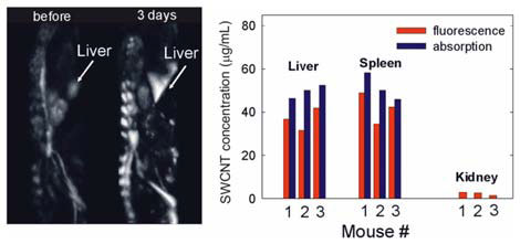 |
| Observed changes in organ brightness in an optoacoustic mouse image can be correlated to quantitative changes in organ absorption coefficients. (© Wiley)
|
|
Usually, due to the unknown light distribution in a complex optical scattering environment, tomographic images of live animals contain only qualitative information and are not suitable for quantitative biodistribution analysis.
|
|
A team of researchers from TomoWave Laboratories, Inc., Rice University, and the University of Houston now developed a methodology to correlate changes in optoacoustic signal intensity from organs of live animals detected with OAT in relation to changes of optical absorption coefficient in those organs caused by nanoparticle accumulation ("Enabling in vivo measurements of nanoparticle concentrations with three-dimensional optoacoustic tomography").
|
|
The researchers quantified localized OAT brightness changes induced by accumulation of single-walled carbon nanotubes (SWCNTs) in liver, kidney and spleen of nude mice. Using the intrinsic fluorescence properties of disaggregated nanotubes, they measured SWCNT concentrations in the parts-per-million range in the harvested organs and defined the corresponding changes in optical absorption coefficient. The observed increases in optoacoustic signal brightness in tissues were compared with the increases in optical absorption coefficients caused by SWCNT accumulation.
|
|
The combination of these methods allows one to perform sensitivity calibration of an OAT system for a selected type of animal and for a range of optical absorption coefficient values of their organs to enable non-invasive concentration measurements of optically absorbing nanoparticles and dyes in vivo.
|