| Posted: Sep 16, 2014 |
Researchers study nanocrystals by passing them through nanopores
|
|
(Nanowerk News) An interdisciplinary team of University of Pennsylvania researchers has now applied a cutting-edge technique for rapid gene sequencing toward measuring other nanoscopic structures. By passing nanoscale spheres and rods through a tiny hole in a membrane, the team was able to measure the electrical properties of those structures’ surfaces.
|
|
Their findings suggest new ways of using this technique, known as “nanopore translocation,” to analyze objects at the smallest scale.
|
|
The research was led by Marija Drndic, professor in the Department of Physics and Astronomy in Penn’s School of Arts & Sciences; Jennifer Lukes, associate professor in the Department of Mechanical Engineering and Applied Mechanics in Penn’s School of Engineering and Applied Science; and Christopher Murray, a Penn Integrates Knowledge Professor who has appointments in both schools through the departments of Chemistry and Materials Science and Engineering. Kimberly Venta, of Drndic’s lab, and Mehdi Bakhshi Zanjani, of Lukes’ lab, were co-lead authors on the paper, and Xingchen Ye and Gopinath Danda also contributed to the work.
|
|
It was published in Nano Letters ("Gold Nanorod Translocations and Charge Measurement through Solid-State Nanopores").
|
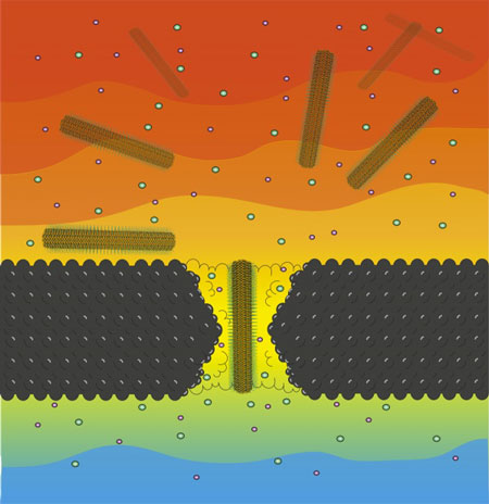 |
| An illustration of a nanocrystal passing through a nanopore.
|
|
For the past several years, Drndic's lab has been exploring an approach to gene sequencing involving DNA translocation through a nanopore. The technique typically involves threading DNA, suspended in an ionic fluid, through a tiny hole in a thin membrane. Each of the four bases of a DNA sequence is expected to block different amounts of the aperture as they pass through, thus allowing a different number of ions to pass through along with them. In most nanopore sequencing, researchers attempt to identify bases by reading changes in the surrounding ion current as it passes through the nanopore.
|
|
This technique has its roots in a device known as a Coulter counter. Such devices have been used for decades to count and sort microscopic particles, like blood cells and bacteria. The principle is the same; particles with larger diameters block more of the aperture, reducing the electrical current measured by electrodes positioned above and below the aperture. This technique has been used on particles that are typically on the micro scale, however, whereas DNA bases are on the nano scale, a thousand times smaller.
|
|
Advances in nanotechnology have allowed researchers to make smaller and smaller pores, and early successes in using this technique with DNA suggested that it could also be applied to better measure other nanoscale structures. Spherical nanocrystals and oblong nanorods, for example, are thought to have potential uses in medicine, electronics and other fields, but their properties must be accurately measured before they can be fine,tuned for their ultimate applications.
|
|
To that end, the members of Drndic’s contingent drew upon their sequencing research involving silicon nitride nanopores, which can be customized to work at various sizes between the nano and micro scales.
|
|
“A great feature of solid-state nanopores is that we can change diameters at will,” Drndic said. “We can use an electron microscope to drill them in whatever size and shape we want, unlike pores in biological membranes, where we would need to find a new system each time.”
|
|
For their measurement targets, the team drew on the Murray lab’s expertise in making uniformly sized gold nanospheres and nanorods that are covered with ligands that give them an overall positive charge. The surface chemistry of these nanoparticles was an attractive match for the translocation technique, which relies on drawing charged objects through the pore.
|
|
“The degree of ligand coverage on the surface of nanoparticles greatly affects the nanoparticle function and quality,” said Murray. “That’s one reason we need to be able to measure them in more detail.”
|
|
The team first used the spherical nanoparticles to calibrate their measurement system.
|
|
“For spherical nanoparticles with charged ligands on their surface,” said Venta, “there is a well-known method for determining the surface charge density, and thus the surface ligand density. However, this method fails for non-spherical nanoparticles.”
|
|
To get around this limitation, the team enlisted modeling expertise from Lukes’ group.
|
|
"Based on the data obtained from the experiments and our computational models," Zanjani said, "we can calculate the surface charge density of the nanorods based on their diameter. Conversely, if we know their surface charge density, we can extrapolate their diameter. The same method can also be used to characterize a variety of other nanoparticles with different sizes and shapes".
|
|
In developing the model for understanding the relationship between these properties, the team also found something unexpected. As nanorods pass through the pore, they typically reduce the ionic current through the pores, as they decrease the amount of space ions can inhabit. However, sometimes an increase in ionic current through pores was recorded.
|
|
|
|
The team determined that this was another area where pore diameter was critical. On average, the pores they drilled were 20 nanometers in diameter, with some a few nanometers wider or narrower. Taking a closer look at these unusual, current-increasing measurements, they determined that, paradoxically, the narrowest pores were triggering them. This suggested that the mechanism had something to do with the proximity between the nanorod and the edge of the pore.
|
|
“There is something about the interaction between the rods and the pores that causes these ‘positive’ events,” said Lukes. “Even though there is less space for the ions to pass through, we think that the current increases because the charged surfaces of the rods and pores attract an even higher concentration of ions than would normally be there for larger pores.”
|
|
This phenomenon could potentially be exploited as different way of measuring particles passing through nanopores. Further research will provide a clearer picture of the diameter tolerances necessary for particles of different shapes. Other aspects of the pore, such as if it has a tapered, hourglass shape versus a smooth, cylindrical one, can also be investigated to see if they make a difference in the kind of signals that can be recorded.
|
|
“This kind of study would not have been possible without Penn’s Materials Science Research and Engineering Center,” Drndic said. “Drawing on physics, chemistry, materials science, mechanical engineering provides us with a unique opportunity to discover interesting phenomena while advancing their practical applications at the same time.”
|