| Posted: Jan 20, 2015 |
A highly sensitive nanoplasmonic biosensor for drug allergy diagnosis
|
|
(Nanowerk News) The new technique is a non-invasive procedure to detect the severity of an allergic reaction to amoxicillin. The developed biosensor platform is based on gold nanodisks, is very sensitive and works label-free, detecting the changes in the refraction index occurring at its surface after the binding of IgEs specific for amoxicillin.
|
|
The bio applications of nanotechnology have an important focus on the development of portable lab-on-a-chip devices. In an article published in Biosensors and Biolectronics ("Highly sensitive dendrimer-based nanoplasmonic biosensor for drug allergy diagnosis"), researchers from the Institut Català de Nanociència i Nanotecnologia (ICN2), in collaboration with University of Málaga and Regional University Hospital of Málaga, have developed a new nanoplasmonic biosensor for drug allergy diagnosis. The ICN2 group in charge of this research is the Nanobiosensors and Bioanalytical Applications Group, led by CSIC Prof Laura M. Lechuga. The first authors of the study are Maria Soler, Pablo Mesa-Antunez and Dr M. Carmen Estevez.
|
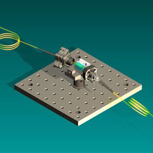 |
| Nanoplasmonic biosensor for drug allergy diagnosis.
|
|
The developed biosensor is able to detect the presence of anti-amoxicillin immunoglobulins E (IgE) in patients’ serum, whose concentration increase during an allergic reaction. Short ordered arrays of gold nanodisks are fabricated on glass substrates. Amoxicillin molecules are immobilized over the gold nanodisks by means of a chemical structure (modified dendrimers) that binds both antibiotic and gold. Finally, the serum of the patient flows over the amoxicillin-coated surface of the sensor.
|
|
The anti-amoxicillin IgEs generated during an allergy outbreak interact with the fixed antibiotic, affecting the properties of the surface. In particular, the binding of the antibody generates a change in the refraction index, which can be monitored. Higher concentration of IgEs will proportionally lead to a higher change in refractive index. With this methodology is therefore possible to quantify the amount IgE in serum, and therefore is possible to diagnose the severity of the allergic reaction.
|
|
This technique stands out because of its high sensitivity, avoiding the use of labels, being possible to detect small amounts of IgEs directly in serum. The biosensing surfaces are excited by a collimated halogen light source at a particular angle, which allows analysing in real time the evolution and changes of gold’s absorption curve during the interaction events. Another outstanding point of this method is the non-invasive procedure for the patient: just few microliters of serum are needed for the analysis.
|
|
The ICN2 researchers have devoted a great effort in the optimization of the methodology, in order to obtain a reproducible and accurate calibration of the biosensor, especially to allow direct detection of serum samples (avoiding pre-treatment and dilution). The strategy has been successfully validated with other established methods like ImmunoCAP. Although this current available technique for allergy diagnosis is well stablished and difficult to replace, the use of a compact and portable platform like this one which minimizes time and costs could mean an attractive alternative for allergy diagnosis, and it could be also very useful in other clinical scenarios.
|
|
At the moment, the biosensor is not available for clinical application as more development is needed to obtain the advanced and quick diagnostic tool they are seeking. However, the aim of the researchers is to design platforms robust and easy to use in order to introduce them in daily clinical practice.
|