| Posted: Oct 01, 2015 |
New DNA stain for live-cell nanoscopy
(Nanowerk News) Ecole Polytechnique Fédérale de Lausanne (EPFL) scientists have developed a new DNA stain that can be used to image living cells.
|
|
One of the holy grails of bio-imaging is to visualize the inner workings of a cell while it is still alive. But this also means that living cells put under the microscope risk being killed by the light and the fluorescent dyes used to highlight their structures. Much like being sunburnt, live cells are sensitive to intense light - and the most dangerous are the ultraviolet and blue lights used in live-cell imaging. EPFL scientists have now developed a new DNA stain that can be used to safely image live mammalian cells for days, even under demanding imaging conditions.
|
|
The work is published in Nature Communications ("SiR–Hoechst is a far-red DNA stain for live-cell nanoscopy"), and it is incorporated into the new EPFL start-up Spirochrome.
|
 |
| Mitosis of a live cell (HeLa) stained with SiR-Hoechst. (Image: Kai Johnsson, EPFL)
|
|
Fluorescent stains that light up a cell's DNA are popular in live-cell imaging as they allow us to track key biological processes such as cell division. However, current DNA stains are themselves toxic or require types of light (e.g. blue) that can damage the cells. Ideally, a safe DNA fluorescent stain would be activated in the safer far-red spectrum of light.
|
|
In addition, many DNA stains are not compatible with super-resolution microscopy, a modern imaging technique that can capture images of cells at higher resolution than that allowed by regular microscopes. Consequently, the bio-imaging community has been waiting for a DNA stain that shows low toxicity, works with far-red light and can be used in superresolution microscopy.
|
|
A DNA stain that does it all
|
|
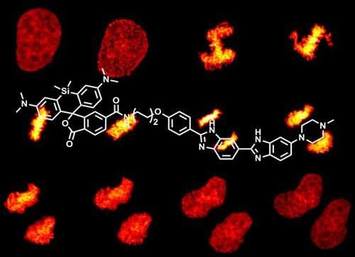
The lab of Kai Johnsson at EPFL has now developed a DNA stain that ticks all the boxes. The scientists combined two molecules. The first is a fluorescent molecule (silicon rhodamine or SiR) that works in the far-red spectrum and was previously developed in Johnsson's lab. The second one is a well-known DNA stain Hoechst (the chemical name is bisbenzimide). As a result, the team named the new DNA stain "SiR-Hoechst".
|
|
The new stain works by binding to a part of the DNA helix known as the "minor groove". Once bound, it turns on and emits a bright fluorescent red light. This is a tremendous advantage, as the stain produces very little noise - if it has not found its target, it stays "off". More importantly, SiR-Hoechst can bind to DNA without affecting its biological function in the cell. And because all cells possess DNA, the probe can be used across numerous species, types of cells, and tissues.
|
|
Because SiR-Hoechst works with far-red light there is little risk of damage to cells. In addition, the light that it emits can be easily distinguished from any background fluorescence of living cells. These features give SiR-Hoechst a clear advantage over other DNA stains: it can safely maintain high-quality staining in live cells for over 24 hours, allowing biologists to identify individual cells in tissue or culture, or track delicate processes, such as cell division, in real time.
|
|
As an additional bonus, the stain can be used in live-cell super-resolution imaging, paving the way for DNA imaging in cells and biological tissues with exquisite resolution. As Kai Johnsson says: "The introduction of SiR-Hoechst brings bioimaging closer to one of its main goals: observing the wonders of nature without disturbing them."
|
|
The team is now preparing to commercialize SiR-Hoechst through their EPFL startup company, Spirochrome. The company has been in business for a while, supplying the scientific community with a new class of fluorescent probes that can image of the cytoskeleton in living cells with unprecedented resolution.
|