| Posted: Jun 18, 2016 |
Dancing platinum particles at atomic resolution
(Nanowerk News) For the first time, near-atomic-resolution movies of individual nanocrystals rotating in solution were used to determine the three-dimensional (3D) atomic structure of nanocrystals. The results yielded insights into the structure and growth mechanisms of these materials (Science, "3D structure of individual nanocrystals in solution by electron microscopy").
|
|
Movies of these nanoparticles in motion were obtained with world-leading electron microscopes using a new graphene-based liquid cell and advanced detectors. The movies were processed with a new theory for image reconstruction to create 3D images.
|
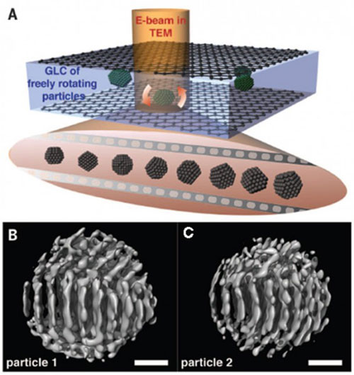 |
| In situ transmission electron microscopy (TEM) imaging of platinum nanoparticles rotating freely in liquid. (A) Schematic representation of individual rotating platinum nanoparticles in a graphene liquid cell (GLC). The bottom represents a series of snapshots of the nanocrystal.(B and C) Two-dimensional snapshots are used to reconstruct the 3D images of two different particles. Scale bars are 0.5 nanometers. (Image: Lawrence Berkeley National Laboratory)
|
|
This new method could further our understanding of how molecular structures migrate and grow in solutions which is critical for the design of more efficient materials for electricity-generating fuel cells, automotive catalytic converters, and other applications.
|
|
Understanding the synthesis, growth, and physical properties of nanoparticles is important for the development of next-generation materials. This research focused on colloidal nanoparticles, clusters of atoms synthesized and suspended in a solution, as many technologically important nano-materials are synthesized this way.
|
|
The size and shape of individual nanoparticles are important for their applications because the properties of the nanoparticles are often controlled by their geometries. Following the growth mechanism or even determining the final 3D structure of nanoparticles is difficult due to the limitations of microscopy.
|
|
A multi-institutional team led by Lawrence Berkeley National Laboratory developed a new technique that provided the first near-atomic-resolution images of colloidal nanoparticles rotating freely in solution. This technique, called Three-Dimensional Structure Identification of Nanoparticles by Graphene Liquid Cell Electron Microscopy, or 3D-SINGLE, combines three technical advances: a vacuum-compatible liquid capsule composed of graphene called a graphene liquid cell, a sensitive detector for imaging, and a computational technique that can reconstruct the high-resolution movies into 3D images.
|
|
The graphene liquid cell shelters the solution from the high vacuum of the transmission electron microscope, and only contains a tiny sample which minimizes unwanted electron beam scattering that causes loss of the atomic resolution.
|
|
The structure of the moving, rotating nanocrystals is imaged many times by a direct electron detector that can produce movies with millisecond frame-to-frame time resolution of the rotating nanocrystals.
|
|
Finally, theoretical advances allow single particle 3D reconstruction. Results confirmed that the individual, and surprisingly asymmetric, particles from the same synthesis solution followed different initiation and growth trajectories.
|
|
Better understanding of synthesis, growth, and properties of individual colloidal nanoparticles could advance the development of novel materials for applications in catalysis and renewable energy.
|