New microscope reveals nanoscale structural dynamics in live cells (w/video)
(Nanowerk News) Scientists at the Marine Biological Laboratory (MBL) and colleagues have unveiled a new microscope that can track the position and orientation of individual molecules in living cells--nanoscale measurements that until now have posed a significant challenge.
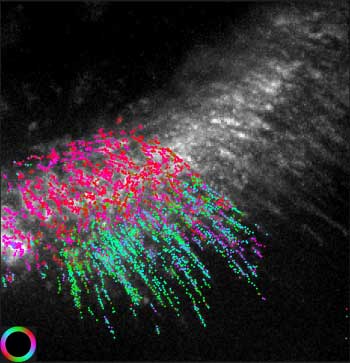
Due to the resolution limit of light microscopes, actin filament orientation in cells was previously accessible only with electron microscopes, but can now be imaged in live cells. Above, color-coded actin filament orientation is overlaid on an image of fluorescence intensity. The orientation corresponds to the color wheel at bottom left. Colored overlay was computed from movies, below. (Image: Shalin Mehta and Tomomi Tani)
"All functions of cells are directed. For example, cells move in a specific direction or divide at a certain site and orientation so the two daughter cells are the right size. That direction comes from the nanoscale alignment of molecules in the cells, which this microscope can detect," says lead author Shalin Mehta, staff scientist in the University of Chicago's Department of Radiology and a staff researcher at MBL.
Understanding how cellular components work requires peering at a nanoscale--to the activity of billionth-of-a-meter-sized molecules that assemble to form the cell's components and drive their functions.
"With this microscope, we can see the orientation of a single molecule, or an assembly of molecules as they form a higher-order structure," says corresponding author Tomomi Tani, an MBL associate scientist. The scope can also detect minute conformational changes that are required for a protein's function.
At left, fluorescent particles move along treadmills of actin filaments in a human skin cell. The images were added along the polarization dimension to compute total fluorescence intensity. At right, orientation of actin filaments at the location of each fluorescent particle. The orientation was computed from polarization-resolved images and is shown by the magenta line. (Video: Shalin Mehta and Tomomi Tani)
Polarized light microscopes, iterations of which been developed at the MBL since the 1950s, exploit "a property of light not visible to the human eye to measure molecular order below the resolution limit of the microscope," Mehta explains.
The team used the microscope to address various biological questions in collaboration with other scientists at the MBL, including Amy Gladfelter of University of North Carolina, Chapel Hill, and Clare Waterman of the National Institutes of Health.
"That is a unique feature of being at the MBL," Mehta says. "We were able to study three biological questions while our method was under development. Trying to solve each question led us to improve the microscope and the algorithms with every iteration."