| Posted: Jul 07, 2017 |
A duet of firsts: Imaging chemical building blocks
(Nanowerk News) Two firsts in science came about because of a near-dare. According to Nigel Browning at Pacific Northwest National Laboratory, “Omar Farha was giving a presentation on MOFs [metal-organic frameworks] and someone said ‘I bet you couldn’t make one out of uranium.’” Farha took the challenge and proved them wrong. In designing the uranium-laden frameworks, PNNL scientists Dr. Nigel Browning and Dr. Layla Mehdi helped Farha and his colleagues at Northwestern University overcome a troubling bottleneck in imaging the material.
|
|
Before this study (Science, "Bottom-up construction of a superstructure in a porous uranium-organic crystal"), scientists used x-ray analysis and modeling to map out MOF structures. The approaches come with sharp drawbacks. Browning and Mehdi showed that low-dose imaging is a viable option for MOF imaging, allowing for the structure to be resolved at the near-atomic level.
|
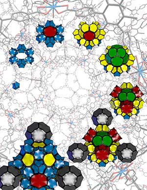 |
| The new metallic-organic framework, NU-1301, is made up of uranium oxide nodes and tricarboxylate organic linkers. (Image: Northwestern University)
|
|
This collaborative effort produced two notable milestones; it was first MOF made out of uranium, and the first time low-dose electron microscopy was used to map the MOF structure.
|
Why It Matters
|
|
The intricate properties of MOFs make them building blocks for chemists. Using the blocks, scientists can design sieves or filters to aid in removing pollutants from different sources or aid catalysts that drive the production of plastics and other goods.
|
|
The problem was that it was incredibly difficult to see the details of the MOFs created in the lab. Previous imaging of MOFs were done using x-ray diffraction, which provides a general overview of the structure, but none of the important details. Models can be used to provide insight on MOF structure; however, they are based off the scientists’ assumptions and can be inaccurate if these assumptions are incorrect.
|
|
“A model is just a basic approximation of what you expect,” says Mehdi, catalysis scientist. “So you’re basically assuming about what you have. And when you have something that drifts away from that model, you have no way to explain that.”
|
Methods
|
|
Scientists shied away from using powerful microscopes to see the structure of the uranium-laden MOF, named NU-1301. A MOF consists of metal ion nodes connected with organic linkers. In NU-1301, the structure is incredibly complex. It consists of 816 uranium nodes and 816 organic linkers. The organic linkers are sensitive to the electron beam, so while electron microscopy would result in a more detailed image, the MOF is damaged or destroyed in the process.
|
|
Browning and Mehdi, along with their team, used low-dose imaging to take images near the atomic scale. With a scanning transmission electron microscopy process, the team directed a dose of one electron per angstrom squared, equal to 0.1 nanometer, in the MOF, allowing for easy imaging of the metal nodes using the Z-contrast imaging mode. Using this method, the arrangement of thousands of uranium rings can be easily seen.
|
|
What’s Next? The next step in the study is to grow the MOF inside of an electron microscope to document how the framework self-assembles. In addition, Browning, Mehdi, and their colleagues will continue to examine the complex structures of NU-1301.
|