| Posted: Sep 08, 2017 |
First imaging of free nanoparticles in laboratory experiment using a high-intensity laser source
(Nanowerk News) In a joint research project, scientists from the Max Born Institute for Nonlinear Optics and Short Pulse Spectroscopy (MBI), the Technische Universität Berlin (TU) and the University of Rostock have managed for the first time to image free nanoparticles in a laboratory experiment using a high-intensity laser source.
|
|
Previously, the structural analysis of these extremely small objects via single-shot diffraction was only possible at large-scale research facilities using so-called XUV and x-ray free electron lasers.
|
|
Their pathbreaking results facilitate the highly-efficient characterisation of the chemical, optical and structural properties of individual nanoparticles and have just been published in Nature Communications ("Coherent diffractive imaging of single helium nanodroplets with a high harmonic generation source").
|
|
The lead author of the publication is junior researcher Dr Daniela Rupp who carried out the project at TU Berlin and is now starting a junior research group at MBI.
|
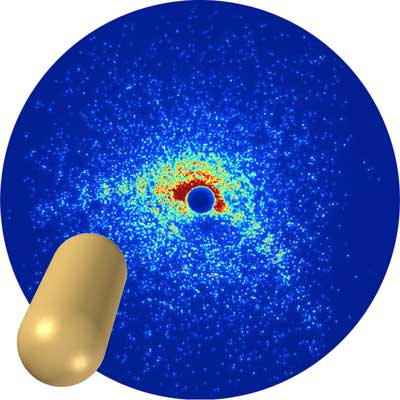 |
| Pill-shaped helium nanodroplets can be detected through curved structures in the scatter image. (Image: MBI)
|
|
In their experiment, the researchers expanded helium gas through a nozzle that is cooled to extremely low temperature. The helium gas turns into a superfluid state and forms a beam of freely flying miniscule nanodroplets.
|
|
“We sent ultra-short XUV pulses onto these tiny droplets and captured snapshots of these objects by recording the scattered laser light on a large-area detector to reconstruct the droplet shape,” explains Dr Daniela Rupp.
|
|
“Key to the successful experiment were the high-intensity XUV pulses generated in MBI’s laser lab that produce detailed scattering patterns with just one single shot,” explains Dr Arnaud Rouzée from MBI.
|
|
“By using the so-called wide-angle mode that provides access to the three-dimensional morphology, we could identify hitherto unobserved shapes of the superfluid droplets,” adds Professor Thomas Fennel from MBI and the University of Rostock.
|
|
The research team’s results enable a new class of metrology for analysing the structure and optical properties of small particles. Thanks to state-of-the-art laser light sources, making images of the tiniest pieces of matter is no longer exclusive to the large-scale research facilities.
|