| Posted: July 3, 2009 |
Novel method of imaging surface charges on individual biomolecules |
|
(Nanowerk News) Surface charges play a key role in determining the structure and function of proteins, DNA and larger biomolecular structures. For example, negatively charged DNA strands electrostatically interact with histone proteins, transcription factors, or polymerases thereby influencing the read-out of genetic information and the development of cancer. Similarly, the central process of protein folding and protein interaction, often governed by charges, is the major factor in protein-folding diseases such as Alzheimer’s or Parkinson’s Disease. However, thus far there have been no experimental methods to spatially resolve the electrostatic surface potential of individual biological molecules. In general, the investigation of individual molecules can shed light on their dynamic behaviour or on static heterogeneity which is masked in ensemble measurements.
|
|
A collaborative effort between researchers from the London Centre for Nanotechnology (Bart W Hoogenboom), King’s College London (Carl Leung, Patrick Mesquida) and UCL Chemistry (Stefan Howorka, Helen Kinns) has led to the first measurements of the electrostatic surface potential of individual DNA and avidin molecules with nanometre resolution using Kelvin Probe Force Microscopy (KPFM) in air (Imaging Surface Charges of Individual Biomolecules).
|
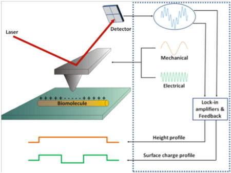 |
| Kelvin Probe Force Microscopy schematic
|
|
Kelvin Probe Force Microscopy (KPFM) can measure surface charges by contactless recording of the electrostatic force between a conductive Atomic Force Microscope tip and a biomolecule on a support. To achieve this, the AFM tip is simultaneously excited at its mechanical resonance frequency and by an electrical (AC) voltage. This periodic electrical voltage on the tip leads to a force between the tip and the charges on the biomolecule, which is recorded by means of a lock-in amplifier and nullified by the Kelvin mode feedback by applying a separate DC voltage (not shown). The polarity and magnitude of this DC voltage corresponds to the local surface charge profile (in mV) which is recorded simultaneously with the topography of the biomolecule.
|
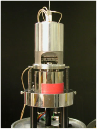 |
| Atomic Force Microscope at the London Centre for Nanotechnology
|
|
The investigation led at the London Centre for Nanotechnology also show, for the first time, the surface potential of buffer salts shielding DNA molecules on a surface, which would not be possible with conventional ensemble techniques. It is anticipated that the ability to visualize the electrostatic surface potentials of individual proteins and DNA at molecular resolution will be an important tool in fundamental biophysical research and in the fields of biosensing and bio-nanoelectronics.
|
|
The London Centre for Nanotechnology, is a UK-based, multidisciplinary research centre forming the bridge between the physical and biomedical sciences. It was conceived from the outset with a management structure allowing for a clear focus on scientific excellence, exploitation and commercialisation. It brings together two world leaders in nanotechnology, namely University College London and Imperial College London, in a unique operating model that accesses the combined skills of multiple departments, including medicine, chemistry, physics, electrical and electronic engineering, biochemical engineering, materials and earth sciences, and two leading technology transfer offices.
|