| Posted: October 23, 2009 |
Free spectral imaging and fluorescence proteins resources on the Zeiss online campus |
|
(Nanowerk News) The educational website for fluorescence microscopy www.zeiss.com/campus has been supplemented with sections on Spectral Imaging and Fluorescent Proteins and now also provides comprehensive information on these topics.
|
|
By visiting the website, interested scientists can learn more about the concepts of spectral imaging and FRET microscopy. The new fluorescent protein section explains how these proteins function and how they are best used in fluorescence microscopy. Furthermore, detailed articles cover topics such as spectral properties, brightness, phototoxicity and photostability of the various fluorescent proteins and offer practical advice on their applications in live cell imaging.
|
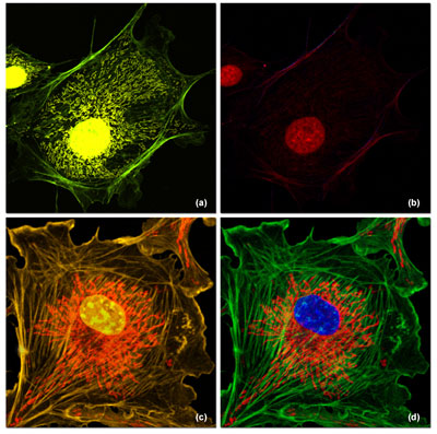 |
| The application photo taken from www.zeiss.com/campus shows examples of spectral imaging and linear unmixing of multiply stained adherent cell cultures with strongly different (a and b) or comparable (c and d) fluorescence intensities.
|
|
The website also comprises interactive tutorials on both topics, e.g. explaining chromophore formation and reactions of fluorescent proteins such as PA-GFP, Kaede and Dronpa or showing how linear unmixing works and FRET biosensors function.
|
|
At www.zeiss.com/campus, users can also find application image galleries. They are invited to share their own results in an application library and they can visit the reference library to find articles on the most important topics in fluorescence microscopy, including spectral imaging, FRET and fluorescence proteins.
|
|
The Online Campus from Carl Zeiss MicroImaging is available as a source of information to scientists and students. Summary reports and interactive Flash animations make it easy for users to quickly learn the various techniques. Theory, technology and applications in fluorescence microscopy are skillfully combined in animations. The website was developed by Mike Davidson, a renowned microscopy expert and online teaching pioneer from Florida State University. Other renowned scientists in the field of fluorescence microscopy already announced that they will make a contribution to the Online Campus.
|