| Posted: March 5, 2007 |
The million dollar microscope |
|
(Nanowerk News) As an inquisitive teenager learning the basics of physics, Stefan Hell believed that the so-called Abbe limit of optical microscopy was an impenetrable barrier. Ernst Abbe, a 19th Century German microscopy pioneer, had declared that resolution beyond half the length of a wave of light - a few hundred nanometers - was impossible.
|
|
'I grew up with this notion,' said Hell, head of nanobiophotonics at the Max Planck Institute for Biophysical Chemistry in Göttingen, Germany. 'It was in all the physics textbooks.'
|
|
But while working toward his PhD, Hell started to become skeptical of Abbe's declaration. 'I got the feeling in 1988 that maybe the last word had not been spoken and I decided to focus on the problem, just for the fun of it.'
|
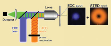 |
| How to break the 'impenetrable barrier' of optical microscopy. (Image: Stefan Hell/Max Planck Institute For Biophysical Chemistry)
|
|
Hell's feeling turned out to be correct. In the years since, he developed the simulated emission depletion (Sted) microscope, which shattered the Abbe limit by producing resolutions of 20 nanometers. German microscope maker Leica Microsystems GmbH, who own exclusive licensing rights to the Sted technology, plan to put the first commercial Sted systems on sale this autumn at about €950,000 (∼ $1.2 million).
|
|
Sted is a cunning extension of a technique called confocal microscopy, where a laser pulse is used to excite fluorescent dye molecules in a sample. Once the dye molecules fluoresce, their light is picked up by a detector and turned into an image, its resolution limited by the size of the laser spot.
|
|
Hell's innovation is to immediately follow the initial pulse, which lasts less than a nanosecond (10-9s), with a second, synchronised laser pulse. The second pulse is redder than the first, tuned to stimulate emission in the dye molecules, and shaped so that it illuminates a ring around the sample rather than a spot.
|
|
Those molecules hit by the body of the ring are forced to dump their energy, while those that sit in the hole in the middle of the ring are allowed to fluoresce as normal. Careful tuning of the second pulse's intensity can narrow the size of the central hole to (in theory) the size of a molecule. This ring-shaped pulse effectively provides a tiny aperture for studying samples. Current state-of-the-art confocal microscopy produces fairly murky images below about 200 nanometers, just where cellular building blocks start to get really interesting. 'This is likely to transform cell biology,' Hell told Chemistry World.
|
|
Focus on single molecules
|
|
Martin Hoppe, global market manager for advanced fluorescent systems at Leica Microsystems, says that Leica has been developing a commercial version of Hell's Sted microscope for four years under the direction of Marcus Dyba, formerly of Hell's Sted team at Max Planck.
The €950,000 list price might sound high, but Hoppe said current top confocal systems that cannot see under 200 nanometers cost as much as €600,000. The extra money for a Sted will make sense for some research groups, he said, and US National Institutes of Health has already expressed interest. Only a limited number of Sted systems will be available in October, and Hoppe said that 'demand will definitely be greater than supply'.
|
|
Stephan Sigrist, of the Rudolf Virchow Center for experimental biomedicine at the University of Würzburg in Germany, who has used a Sted prototype, described it as a 'really groundbreaking innovation' that will give scientists a real-time view of molecular architecture never before seen in cells.
|
|
Diego Restrepo, director of the Neuroscience Program at the University of Colorado at Denver and Health Sciences Center (UCDHSC), used a Sted microscope prototype in Hell's lab in Göttingen. 'This was a once in a lifetime experience,' he said. Using the Sted, Restrepo and his team imaged samples of olfactory epithelium, nasal tissue that is involved in smell. The cells were stained with a fluorescent indicator for an ion receptor channel, TRPM5. The results show that TRPM5 is expressed in small clusters of 70 nm diameter in the cilia of the olfactory sensory neurons.
|
|
'It is not much of an exaggeration to say that every cell biologist in the world will have a research project of interest to implement with the Sted microscope,' he said, adding that a group of fellow UCDHSC biomedical scientists has already applied to the National Science Foundation for funding to purchase a Leica Sted microscope.
|
|
Asked whether it is possible to develop optical microscopes capable of producing an image of a single molecule, Hell said: 'I think it is possible. I think in the future we will do this.'
|