| Posted: July 6, 2010 |
Multicolor quantum dots aid in cancer biopsy diagnosis |
|
(Nanowerk News) The tunable fluorescent nanoparticles known as quantum dots make ideal tools for distinguishing and identifying rare cancer cells in tissue biopsies, Emory and Georgia Tech scientists have demonstrated.
|
|
An article to be featured on the cover of the July 15 issue of Analytical Chemistry ("Multiplexed Detection and Characterization of Rare Tumor Cells in Hodgkin's Lymphoma with Multicolor Quantum Dots") describes how multicolor quantum dots linked to antibodies can distinguish the Reed-Sternberg cells that are characteristic of Hodgkin's lymphoma.
|
|
"Our multicolor quantum dot staining method provides rapid detection and identification of rare malignant cells from heterogenous tissue specimens," says senior author Shuming Nie, PhD, the Wallace H. Coulter distinguished professor in the Coulter department of biomedical engineering at Georgia Tech and Emory University. "The clinical utility is not limited to Hodgkin's lymphoma but potentially could be extended to detect cancer stem cells, tumor-associated macrophages and other rare cell types."
|
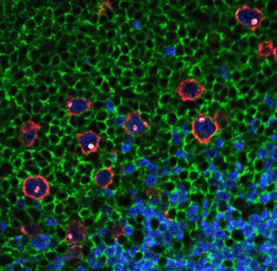 |
| Reed-Sternberg cells can be distinguished by their red outline, blue and white internal staining, and their lack of green staining.
|
|
Quantum dots are nanometer-sized semiconductor crystals that have unique chemical and physical properties due to their size and their highly compact structure. Quantum dots can be chemically linked to antibodies, which can detect molecules present on the surfaces or internal parts of cancer cells.
|
|
As a test of quantum dots' discriminatory power, the authors used four varieties at once -- white, red, green and blue – each detecting a different protein, to stain lymph node biopsies. The goal was to distinguish six Hodgkin's lymphoma cases from two other types of lymphoma and samples from two patients with benign growths in their lymph nodes.
|
|
Reed Sternberg cells have a distinctive appearance, but in lymph node tissue, they are usually surrounded by other white blood cells. The authors describe identifying them as a task like "finding a needle in a haystack."
|
|
"We're excited about this technology," says Andrew Young, MD, PhD, associate professor of pathology and laboratory medicine at Emory University School of Medicine and director of clinical laboratories at Grady Health System. "We expect it could help guide the type of treatment a cancer patient gets and that it could be used with a wider variety of tumor types."
|
|
The most reliable way to assign cell identity is to look at more than one protein, Young says. With the standard methods in most pathology labs, staining cells with four different antibodies would require four separate slides – a problem when the specimen is very small. Small diagnostic specimens are common today, because they minimize the burden on the patient. In addition, the images from multiple separate slides wouldn't depict exactly the same cells. The quantum dots allow "multiplexing": superimposing four colors on top of each other.
|
|
Hodgkin's lymphoma is usually treated with chemotherapy and radiation, and is notable among the subtypes of adult lymphoma because the survival rate is relatively high. Young says the quantum dot technique could be useful for other types of cancer, where distinguishing cancer cells based on surface or genetic markers can point oncologists towards "targeted therapies" designed for one particular type of tumor.
|