| Jan 12, 2011 |
Flares on the move: Nanoparticle test kit shows how nanoparticles of different size disperse in tumor tissue
|
|
(Nanowerk News) Nanoparticles play a significant role in the development of future diagnostic and therapeutic techniques for tumors, for example as transporters for drugs or as contrast agents. Absorption and dispersion of nanoparticles in tumor tissue depend strongly on particle size. In order to systematically study this, scientists at the Massachusetts Institute of Technology and Harvard Medical School have now produced a set of fluorescent nanoparticles of various diameters between 10 and 150 nm. As the team led by Moungi G. Bawendi and Daniel G. Nocera reports in the journal Angewandte Chemie ("A Nanoparticle Size Series for In Vivo Fluorescence Imaging"), they were able to use these to simultaneously follow the dispersion of particles of different sizes through mouse tumors in real time.
|
 |
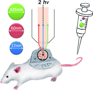
| A nanoparticle toolset was created within the size limits of 10-150 nm for probing size-dependent nanoparticle distribution in solid tumors. By using multiphoton intravital microscopy, the particles were tracked both spatially and temporally in the same tumor grown in a transparent window model
|
|
In order for nanoparticle-based biomedical techniques to work, the nanoparticles must be of optimal size. For studies, it is thus desirable to simultaneously observe the behavior of particles of different size in the same tumor in vivo. This requires chemically comparable particles of various sizes, each size group consisting of particles of uniform size and composition. Additionally, it must be possible to simultaneously detect and differentiate the various particles. Also, they must be biocompatible, and may not form aggregates or adsorb proteins. This complex challenge has now been met.
|
|
The researchers developed a set of nanoparticles in various sizes, which can be detected by means of fluorescing quantum dots. Quantum dots are semiconducting structures at the boundary between macroscopic solid bodies and the quantum-mechanical nano-world. By selectively producing quantum dots of different sizes, it is possible to obtain quantum dots that fluoresce at different defined wavelengths, which allows them to be simultaneously detected and differentiated.
|
|
To produce nanoparticles in different size classes, the scientists coated cadmium selenide/cadmium sulfide quantum dots with polymer ligands such as silicon dioxide and polyethylene glycol. They attained particles larger than 100 nm in diameter by attaching quantum dots to prefabricated silicon dioxide particles and then coating them with polyethylene glycol. For each size class they selected quantum dots that give off light of a different wavelength.
|
|
The researchers intravenously injected a mixture of particles with diameters of 12, 60, and 125 nm into mice with cancer. Fluorescence microscopy was used to follow the particles' entry into the tumor tissue in vivo. Whereas the 12 nm particles easily passed from the blood vessels into the tissue and rapidly spread out, the 60 nm particles passed through the walls of the vein but stayed within 10 µm of the vessel wall, unable to pass farther into the tissue. The 125 nm particles essentially did not pass through the walls of the blood vessels at all.
|

