New microscope produces dazzling 3D movies of live cells
(Nanowerk News) A new microscope invented by scientists at Howard Hughes Medical Institute's Janelia Farm Research Campus will let researchers use an exquisitely thin sheet of light -- similar to that used in supermarket bar-code scanners -- to peer inside single living cells, revealing the three-dimensional shapes of cellular landmarks in unprecedented detail. The microscopy technique images at high speed, so researchers can create dazzling movies that make biological processes, such as cell division, come alive.
The technique, called Bessel beam plane illumination microscopy, is described in a research article published online on March 4, 2011, in the journal Nature Methods.
High-speed imaging with the Bessel beam plane illumination microscope reveals the ever-changing surface of a HeLa cell, with long, thin projections called filopodia continually extending and retracting. (Video: Laboratory of Eric Betzig/Janelia Farm)
A major goal of biologists is to understand the rules that control molecular processes inside a cell. If one is trying to learn the rules of a game, it is better to have a movie of people playing the game than it is to have still photos — and the same is true for cells, says Janelia Farm group leader Eric Betzig. He has been inventing and improving microscopes for more than 30 years. Despite having seen huge advances in microscopy during that time, Betzig says the field is still hindered by the fact that many microscopy techniques require that cells be killed and fixed in position for imaging. There is only so much one can learn from studying dead cells – the equivalent of still photos, he says.
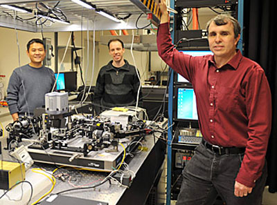
Liang Gao, Thomas Planchon and Eric Betzig display their new Bessel beam plane illumination microscope at HHMI's Janelia Farm Research Campus.
Betzig wanted to create a microscope that would let researchers see the dynamic inner lives of living cells. The notion of studying live cells, stippled with fluorescently labeled proteins and other molecules, is not new. But live-cell techniques can be problematic because light produced by microscopes can damage the cell over time. Besides cell damage, light causes the fluorescent molecules --of which there are only so many -- to wink out over time. In other words, the longer you study the cell to uncover its properties, the more damage you do to the cell and the more likely you are to spend your "photon budget," Betzig says.