| Mar 23, 2011 |
Contrast agent for tumor diagnostics
|
|
(Nanowerk News) X-rays are not the only way: visible and especially infrared light can also be used to image human tissue. The effectiveness of optical imaging processes can be significantly improved with suitable dyes used as contrast agents.
|
|
In the journal Angewandte Chemie ("Phosphorescent Nanoscale Coordination Polymers as Contrast Agents for Optical Imaging"), a team led by Wenbin Lin at the University of North Carolina, Chapel Hill, USA, has now introduced a novel contrast agent that marks tumor cells in vitro. The dye is a phosphorescent ruthenium complex incorporated into nanoparticles of a metal–organic coordination polymer, which allows an extraordinarily high level of dye loading.
|
|
Fluorescent dyes accumulate in varying amounts in different types of tissue. Such contrast agents make it possible to use optical imaging to differentiate between healthy and tumorous tissue. However, this method is limited by the fact that very high concentrations of dye are needed to produce sufficiently strong fluorescence. Organic dye molecules packed at high concentrations into nanocapsules tend to quench each other's fluorescence. Materials that fluoresce more strongly, such as quantum dots, are often not biocompatible.
|
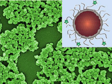 |
| Phosphorescent nanoscale coordination polymers (NCPs) with unprecedentedly high dye loadings were coated with thin silica shells to tune the dye release kinetics (see picture). Further functionalization of the NCP/silica particles with poly(ethylene glycol) (PEG) and PEG-anisamide enhanced their biocompatibility and targeting ability, allowing cancer-specific imaging of human lung cancer H460 cells.
|
|
Metal and Polymer Lattices
|
|
This team has now developed an alternative: metal complexes connected to form lattice-like coordination polymers. Coordination polymers are metal–organic structures consisting of metal ions, which act as connecting points, linked by bridges made of organic molecules or coordination complexes. The scientists made such polymers with bridges consisting of a light-emitting complex of the metal ruthenium. Zirconium ions proved to be suitable connecting points. These tiny structures form spherical nanoparticles.
|
|
The ruthenium complexes do not fluoresce, but rather phosphoresce, which means that they emit light for a proportional length of time after irradiation with light. Because they are not placed inside a nano-transport container, but are a component of the nanoparticle, it is possible to attain a very high level of dye loading—in this instance over 50 %. Quenching of the phosphorescence at high concentrations does not occur in such complexes.
|
|
Increasing Biocompatibility
|
|
In order to prevent the glowing particles from rapidly dissolving and to increase the biocompatibility, they were coated with thin layers of silicon dioxide and a layer of polyethylene glycol. The latter acts as an anchor point for anisamide, a molecule that specifically binds to receptors that are far more common on the surfaces of many types of tumor cell than on healthy cells.
|
|
In a cell culture, it was possible to selectively mark a line of cancer cells with the phosphorescent nanoparticles. The researchers hope that it will be possible to develop contrast agents for the use of optical imaging for tumor detection based on these new metal–organic nanomaterials.
|