| Jun 22, 2011 |
Optics: Through the crystal spheres
|
|
(Nanowerk News) One of the oldest and most important scientific instruments ever invented is the optical microscope. Some people use microscopes to examine small animals, such as flies and ants, while others use them to study even tinier objects, such as cells and tissues. Due to diffraction, however, the highest resolution that an optical microscope can achieve has been limited to a few hundred nanometers. Now, Boris Luk'yanchuk at the A*STAR's Data Storage Institute and co-workers have discovered a simple way to push the resolution of an optical microscope beyond the diffraction limit. Using micrometer-sized quartz spheres, they have demonstrated the possibility of imaging down to 50 nanometers ("Optical virtual imaging at 50 nm lateral resolution with a white-light nanoscope").
|
|
According to classical optic theory, the resolution of an optical instrument is limited to about half the wavelength of light used, which is about 200 nanometers for visible light. This limit can be enhanced through the use of 'superlenses' made of metamaterials, which do not obey the same laws of optics as conventional materials. However, metamaterials are difficult to fabricate, not very efficient, and provide their superlensing functionality only for a very narrow range of wavelengths.
|
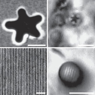 |
| Scanning electron microscopy images (left) of objects with sub-100 nanometer features (scale bar: 500 nm), and microsphere-enhanced optical microscopy images (right) of the same features (scale bar: 5 ?m), demonstrating resolution beyond the optical limit.
|
|
Luk'yanchuk and his fellow researchers developed a more practical approach that involves the use of tiny quartz spheres. It is well-known that quartz spheres can image and focus light—an effect that can be readily observed using glass marbles. However, at the microscale, the lenses image and focus light not through refraction, but through the scattering of light. The quartz microspheres, each 2–9 micrometers in diameter, are inserted in a precise geometry between the lens and the object. In the present case, this allowed the researchers to achieve a magnification enhancement of four to eight times.
|
|
In comparison with superlenses, the benefit of these spheres is that they are easy to fabricate and that they can be used across the entire visible spectrum. The smallest structures that could be resolved this way is 50 nanometers (see figure), far beyond the diffraction limit of regular lenses.
|
|
This microsphere-based approach may have a broad range of applications, particularly in biotechnology, where optical microscopes are regularly used. "For example, it is not possible to observe live viruses by electron microscopy, but it is possible with this super-enhanced optical microscope," says Luk'yanchuk. There are still issues to be resolved, but Luk'yanchuk is optimistic that sooner or later their technology will be used across all optical microscopes. "History tells us that once we have solved the basic physics, the technology will follow." |