| Jun 27, 2011 |
Fluorescent diamond nanocrystals offer new possibilities for biosensing in living cells
|
|
(Nanowerk News) The real-time imaging of dynamic processes within living cells is crucial in order to understand biological functions in detail. This is particularly important in the field of targeted drug delivery, which relies on specific interactions of nanoparticles inside cells. Fluorescence imaging using green fluorescent protein (GFP) is a commonly used technique for observing interactions in live cells, but the GFP approach cannot be used to track the position and orientation of individual molecules or nanoparticles.
|
|
Now, researchers from the University of Melbourne in Australia have developed a diamond nanoparticle-based method that allows the position and orientation of the fluorescent particles to be monitored in living cells ("Quantum measurement and orientation tracking of fluorescent nanodiamonds inside living cells").
|
|
"Diamond is a proven highly biocompatible material that coupled with its magnetic properties and long-term stability of its fluorescence serves as a powerful tool for biomarker applications," says Lloyd Hollenberg from the research team.
|
 |
| Fluorescence imaging of nanodiamonds in a living human cell. (© 2011 NPG)
|
|
Diamond's long-lived fluorescence, which persists for as long as 10 hours, arises from nitrogen impurities known as 'NV' centers in its crystal structure. Most promising for biosensing applications, the orientation of the magnetic spin of an atomic-scale NV center can be monitored using an optical microscope.
|
|
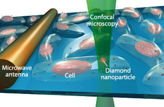
In their study, the researchers inserted diamond nanocrystals as small as 50 nm into living human cells in culture. They then excited the NV centers into specific overall spin states using microwave radiation emitted from an antenna held close to the cell, and monitored the orientation of the NV spin from the fluorescence of the nanocrystals upon momentary excitation with a laser (see image). Hollenberg and his team found that each nanocrystal has its own unique spectral fingerprint, which makes tracking the movement of individual nanocrystals through the cell a straightforward task. The technique also provides information on a property of the nanoparticles called quantum coherence, which could open up new insights into intracellular processes.
|
|
This diamond nanoparticle-based imaging technique has many possible applications, says Hollenberg. "The ability to fingerprint the atomic levels and track both the position and orientation of individual nanoparticles within a cell over long periods of time could provide information of interest in nanomedicine, where the uptake and trafficking of nanoparticles is critical to successful drug delivery targeting."
|