| Jun 28, 2011 |
New method for imaging molecules inside cells
|
|
(Nanowerk News) Using a new sample holder, researchers at the University of Gothenburg, Sweden, have further developed a new method for imaging individual cells ("An in situ fracture device to image lipids in single cells using TOF SIMS"). This makes it possible to produce snapshots that not only show the outline of the cell's contours but also the various molecules inside or on the surface of the cell, and exactly where they are located, something which is impossible with a normal microscope.
|
|
Individual human cells are small, just one or two hundredths of a millimetre in diameter. As such, special measuring equipment is needed to distinguish the various parts inside the cell. Researchers generally use a microscope that magnifies the cell and shows its contours outline, but does not provide any information on the molecules inside the cell and on its surface.
|
|
"The new sample holder is filled with holds cells in solution," says Ingela Lanekoff, one of the researchers who developed the new method at the University of Gothenburg's Department of Chemistry. "We then rapidly freeze the sample down to -196°C, which enables us to get a snapshot of where the various molecules are at the moment of freezing. Using this technique we can produce images that show not only the outline of the cell's contours, but also the molecules that are there, and where they are located."
|
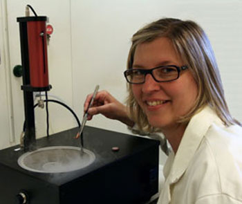 |
| Ingela Lanekoff with a cell sample in the new sample holder.
|
|
So why do the researchers want to know which molecules are to be found in a single cell? Because the cell is the smallest living component there is, and the chemical processes that take place here play a major role in how the cell functions in our body. For example, our brain has special cells that can communicate with each other through chemical signals. This vital communication has been shown to be dependent on the molecules in the cell's membrane.
|
|
Imaging the molecules in the membrane of single individual cells's membrane enables researchers to measure changes. Together with previous results, Lanekoff's findings show that the rate of communication in the studied cells studied is affected by a change of less than one per cent in the quantities abundance of a specific molecule in the membrane. This would suggest that communication between the cells in the brain is heavily dependent on the chemical composition of the membrane of each individual cell,. This could be an important part of the puzzle which could go some way towards explaining the mechanisms behind learning and memory.
|
|
The thesis also describes a new method whereby specific molecules are used in combination with special measuring equipment to locate bacteria that live on the seabed oceanfloor. The bacteria in question play an important role in nature as they counteract both seabed oceanfloor death and eutrophication overfertilization. The method enables researchers to monitor the depth and location of these bacteria in sediment on the seabed oceanfloor.
|