| Posted: July 11, 2007 |
Big improvement for little pictures |
|
(Nanowerk News) Researchers who want to monitor changing nanoscale structures in real time often must do without depth information. But in the 6 July Physical Review Letters ("Imaging Surface Topography using Lloyd's Mirror in Photoemission Electron Microscopy"), physicists describe a new way to view three-dimensional structures in real time. Although they haven't yet published 3-D movies, they managed to reconstruct the heights of tiny droplets using still images from 2-D movies. The technique could give researchers a way to watch technological processes like semiconductor growth as they happen.
|
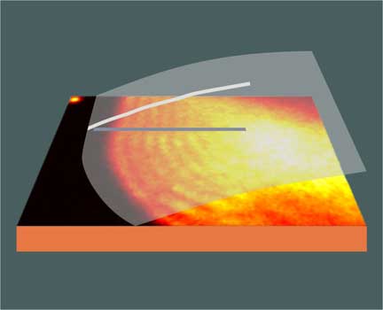 |
Lights, nanocamera, action! Light and dark bands in a flat image of a gallium droplet can lead to the vertical profile of the droplet (white line) and the extrapolated droplet surface (transparent white). This technique may allow researchers to make high-speed, three-dimensional movies of nanostructures. (Image: D. Jesson/Monash University)
|
|
Techniques such as atomic-force microscopy and scanning tunneling microscopy let researchers map three-dimensional samples in exquisite detail. But these methods are too slow to capture fast-moving events because they build up images by sequentially measuring the height at each point on a surface. A method called photoemission electron microscopy, or PEEM, solves the speed problem for two-dimensional images by shining high-energy light on an entire sample. Electrons ejected by the light are focused to create a video of a large portion of the sample at once. Unlike other techniques, a PEEM movie can show a growing crystal, an oscillating droplet, or shifting magnetic domains. Until now, however, the technique couldn't show how different regions compare in height.
|
|
David Jesson and his team at Monash University in Australia used PEEM to investigate droplets of liquid gallium sitting on a mirror-flat substrate of gallium arsenide, like raindrops on a the hood of a car. When they looked closely at the images, they noticed bright and dark bands tracing the boundaries of the droplets. "It was something that was very unexpected, and we figured we should really sort it out," says Jesson.
|
|
Luckily, he remembered a century-old optical effect called Lloyd’s Mirror, in which light shining at a mirror interferes with light reflected from it. In his PEEM experiment, the light comes in at a small angle to the surface, some of it bouncing off the reflective gallium arsenide before hitting a nearby droplet. This light interferes with light that hits the droplet directly. The interference generates parallel light and dark lines of illumination wherever the sample height changes, which leads to light and dark lines in the electron image.
|
|
The bright and dark bands, or "fringes," resemble the lines on a topographic map that indicate varying altitude. The researchers used them to determine the three-dimensional shape of the droplets and could potentially watch the droplets as they join into wider and taller structures. "All we have to do, in principle, is count the fringes to get the height," says Jesson. Other PEEM researchers have noticed the fringes, but until now they weren’t considered useful. "I suspect they regarded them as a nuisance," he says.
|
|
Jesson’s group used a laboratory ultraviolet lamp, which lets them see surface features no smaller than several hundred nanometers tall. The team expects to improve this resolution to tens of nanometers by using light from a synchrotron, a large facility dedicated to research with light beams. Under ideal conditions, they might be able to catch action on the hundred picosecond time-scale, limited only by the speed of the digitizing equipment.
|
|
According to Jesson, nanoscale PEEM movies might help researchers study the quick magnetic changes in nanostructures that are being developed for instant-on computer memory. They may also be useful for controlling the growth of nanostructures and crystals.
|
|
Hendrik Ohldag of Stanford University in California uses synchrotron PEEM to examine magnetic memory materials. "Any kind of depth information is very valuable," says Ohldag. "It would be nice to see the extension of the [magnetic] domain perpendicular to the surface."
|