| Aug 09, 2011 |
Better batteries through nanoscale 3D imaging
|
|
(Nanowerk News) As an important step toward reducing oil dependence and greenhouse gas production, electric vehicles are becoming more and more prevalent. However, one major barrier remains: their batteries. Today's lithium-ion technology has yet to meet energy density, cost, life cycle and safety goals.
|
|
To combat these difficulties, two recent studies conducted at SSRL Beam Line 6-2 used a combination of techniques, together termed "X-ray Absorption Near-Edge Structure (XANES) Microscopy," to study lithium-ion batteries. XANES Microscopy, with its ability to combine spatial and energy resolution over large areas with fast acquisition, is likely to benefit not only the field of energy storage, but also fields ranging from biomaterials to archaeology.
|
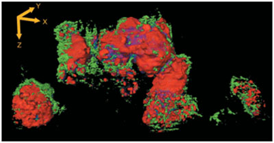 |
| Reconstructed three-dimensional XANES tomography data of a Li-ion battery electrode.
|
|
The first of the two studies, led by Wilson Chiu from the University of Connecticut, obtained nanoscale spatial information about materials found in lithium-ion battery electrodes. The second study, led by Fondazione Bruno Kessler's Florian Meirer and Lawrence Berkeley National Laboratory's Jordi Cabana, looked at the battery electrodes themselves. The studies provided 2D and 3D chemical information about the changes taking place in the electrodes, revealing the location of nickel and nickel oxide. By correlating this with changes in the morphology and porosity of the cathode, this work provides a new perspective on lithium-ion battery electrodes that could direct new design strategies for the next generation of batteries.
|
|
These studies were published in Applied Physics Letters ("Three-dimensional mapping of nickel oxidation states using full field x-ray absorption near edge structure nanotomography") and the Journal of Synchrotron Radiation ("Three-dimensional imaging of chemical phase transformations at the nanoscale with full-field transmission X-ray microscopy"), respectively.
|