| Jan 24, 2006 |
Ultrafast nanolaser flow device for detecting cancer in single cells
|
|
(Nanowerk News) To investigate tumors, pathologists
currently rely on labor-intensive microscopic examination,
using century-old cell-staining methods that can take days
to complete and may give false readings.
|
|
A lightning-fast laser technique, led by Sandia National
Laboratories researcher Paul Gourley, has provided laboratory
demonstrations of accurate, real-time, high-throughput identification
of liver tumor cells at their earliest stages, and without
invasive chemical reagents. The work is detailed in the December 2005 issue of the journal Biomedical Microdevices.
|
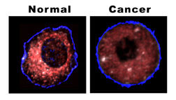 |
Proof positive — The difference between a normal and cancerous liver cell is shown clearly by the location of mitochondria, as revealed by Sandia's biocavity laser. The healthy cell shows very few mitochondria near the outer cell wall; they cluster densely (red coloration) as they approach the cell's nucleous (depicted here as the black central hole). In the cancerous cell, the mitochondria are spread throughout the cell, do not cluster, and under the same lighting produce a more subdued effect.(Source: Sandia National Laboratories) |
|
The technique generates a laser beam in single human cells
pumped from a flask through tiny microchannels. The beam
is altered by what it encounters. These changes, registered
by an imaging spectrometer, instantly identify cancer-modified
mitochondria in cells gone wrong. Mitochondria are known
as the power pack of cells, energizing them like batteries
do flashlight bulbs.
|
|
“There are hundreds of mitochondria, sometimes thousands,
in a cell,” says Gourley. “To see them in the
old way requires a time-consuming process like fluorescent
tagging or a chemical reagent. We’ve found we can
see them immediately by light alone.”
|
|
The techniques could be critical to advancing early detection,
diagnosis, and treatment of disease.
|
|
More technically put, “To rapidly assess the health
of a single mammalian cell,” says Gourley, “the
key discovery was the elucidation of biophotonic differences
in normal and cancer cells by using intracellular mitochondria
as biomarkers for disease. This technique holds promise
for detecting cancer at a very early stage and could nearly
eliminate delays in diagnosis and treatment.”
|
|
The technique is effective because “it measures changes
in the cell architecture, especially those arising from
alterations in protein density, cytoskeleton shape, and
distribution of mitochondria — changes that occur
when a cell becomes cancerous,” says Gourley.
|
|
“One would think that if a cell became nonfunctional,
it would become disorganized. In cancer, however, that’s
not the case. A cancer cell is like an insurgent terrorist
with a very well-defined agenda. It rearranges the cytoskeleton
and the arrangement of mitochondria in the cell. It’s
no longer a cooperative agent in a collection of cells but
becomes malicious, tries to get outside the area, and hijacks
the respiratory machinery of a cell.”
|
The biocavity laser
It is these changes — a kind of beefing-up of the
criminal forces — that Gourley’s device, called
a biocavity laser, detects.
|
|
A nano-thin layering of gallium aluminum arsenide combinations
send up numerous tiny beams from a small cross-sectional
generating area. These beams are reinforced or thwarted
by the position and density of the mitochondria.
|
|
“The pictures we get from normal and cancer cells
are very different,” says Gourley. “Mitochondria
conspire to cluster around the nucleus and work together
to supply energy to the healthy, functioning cell. In contrast,
the mitochondria in the cancer cell sit all over, isolated
and balled up in a quiescent, non-functioning state. Apparently,
the rapidly growing cancer cells derive energy from an alternative
source such as free glucose in the cell.”
|
|
Fortunately, the mitochondrion is nearly the same size
as the light wavelength of about 800 nanometers, a frequency
otherwise little absorbed by the body. Because of this close
match, the laser is exquisitely sensitive to subtle changes
in the mitochondria size and effects of clustering. To date,
the research team has found that 90 to 95 percent of light
scatter generated is from optical properties of mitochondria.
|