| Feb 22, 2006 |
Researchers in Germany unveil the first three-dimensional electron microscope for examining nanomaterial structures
|
|
(Nanowerk News) The world’s first electron microscope for simultaneously and automatically investigating in three-dimensions the phase content,
crystallographic texture, and crystal interfaces of materials was co-designed and put into service at the Max Planck Institute for Iron Research in Düsseldorf, Germany.
|
|
The approach, called "3D EBSD-FIB technique", is realized by a combination of a focused ion beam (FIB) unit for serial sectioning with high-resolution field emission scanning electron microscopy with electron backscattering diffraction (EBSD) in one device. In the past, these two types of microscopes have been used separately; now, they have been integrated into a single instrument together with an arsenal of detectors which can measure electron diffraction patterns and orientations, as well as perform chemical analyses. This allows scientists to see the inner structure of nanomaterials, biological matter, and high-performance steels in ways that other microscopic procedures cannot - and in full 3D.
|
|
The researchers report their findings in the article "Investigation of orientation gradients around a hard Laves particle in a warm-rolled Fe3Al-based alloy using a 3D EBSD-FIB technique" in the Jan. 3, 2006 online edition of Acta Materialia.
|
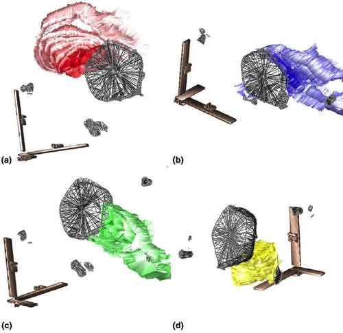 |
| 3D visualization of the four orientation gradient zones in the crystallographic orientations in an intermetallic iron-aluminium specimen (lattice curvature) close to a very hard Laves phase (appears as a transparent net). The various layers of colour indicate increasing changes in the crystal orientation from the reference point directly at the interface between matrix and Laves phase in two degree
misorientation intervals. A characteristic color code is used for each separate gradient zone: (a) gradient 1; (b) gradient 2; (c) gradient 3; (d) gradient 4. (Source: Max Planck Institute for Iron Research; Reprinted from Acta Materialia, 54 (2006), 1369–1380, J. Konrad et al., with permission from
Elsevier)
|
|
The chance to investigate microstructures three-dimensionally is of great use to materials scientists. Until
now, there have been two ways to make such observations. The first is employing x-rays or electron or
neutron beams, respectively. The advantages of these methods are that it they are not destructive and let
scientists reconstruct the three-dimensional structure. But this process is also time-consuming and provides
only limited information - particularly for crystalline materials. Also, the lateral resolution is about two
orders of magnitude below that of the new 3D-approach the Max Planck scientists have now unveiled, which
reaches at least 40 cubic nanometres.
|
|
The second option has been to take an image of the material in slices, and then reconstruct it tomographically
in three-dimensions. The new microscope makes three-dimensional images by first taking a two-dimensional
picture using the desired crystallographic or chemical method. Then, with nanoscopic precision, an ion beam
slices off a piece of the material - and the layer underneath can be analyzed. The researcher can then
investigate the material slice-for-slice, and in the end, the device produces a digital three-dimensional image.
|
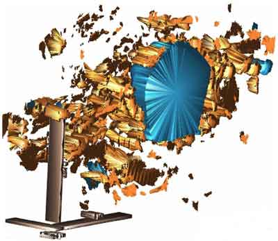 |
| 3D visualization of areas of low local misorientation in the vicinity of the Laves-phase particle. The Laves phase is colored blue. The areas which are characterized by a weak average local misorientation of less than 2.5 are colored gold.. (Source: Max Planck Institute for Iron Research; Reprinted from Acta Materialia, 54 (2006), 1369–1380, J. Konrad et al., with permission from Elsevier)
|
|
The microscope functions fully automatically. This allows relatively large areas to be investigated - for
example, volumes of 70 x 70 x 70 micrometres. The powerful combination of automatically slicing off
material and looking at it in high resolution produces a range of crystallographic information much broader
than most other microscopy techniques. That information includes the exact form of the embedded crystals,
the position and crystallographic characteristics of internal interfaces, the density of defects in grains, and
very fine textural details. All these characteristics can be measured at a lateral resolution of about 40 cubic
nanometres - and depending on the material, even more finely.
|
|
Max Planck researchers working with Prof. Dierk Raabe and Dr. Stefan Zaefferer have already looked at
steel-related iron-aluminium intermetallic alloys. These alloys are characteristically highly resistant to
oxidation and sulphidation at high temperatures. On the other hand, they are not yet completely hard and
homogenous. For this reason, the scientists are adding tiny particles and chromium, in an attempt to refine
the materials for technical application. Such alloys could be used in newly-designed high-temperature gas
turbines for conventional power plants, allowing plants to operate more efficiently and ecologically, and
bringing down energy costs.
|
|
In the case of the iron-aluminium intermetallic alloy, Raabe and his colleagues are using the instrument to
look at the effect of embedded particles on the microstructure and macroscopic characteristics of the material.
In particular, the scientists investigated how tiny inclusions in the intermetallic matrix influence the
orientation of the alloy’s crystal lattice.
|
|
In hot-formed samples the scientists were able to observe for the first time in 3D how soft crystal orientations
of the matrix developed substantial orientation gradients around hard particles. The crystallographic
orientations took on specific patterns, characterised by the building up of systematic orientation gradients
in a number of successive layers which had increasing orientation distance from the interface with the hard
inclusion. Because of these strong crystallographic gradients, new seed crystals developed, homogenising
the material and improving its mechanical characteristics in view of high-temperature application in modern
power plants.
|