| Posted: March 13, 2008 |
Wellcome Image Awards 2008 |
|
(Nanowerk News) Bold, beautiful and groundbreaking, this year's Wellcome Image Awards delve deep into our understanding of modern medicine and science.
|
|
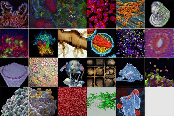
Dazzling and brightly coloured images depict subject matters as diverse as breast cancer cells, Clostridium difficile and ruptured blood vessels, and exemplify recent advances in science and medicine.
|
 |
|
Each year, the Wellcome Trust runs the Wellcome Image Awards ceremony (previously the Biomedical Image Awards) to recognise the scientists who have created stunning and beautiful images as part of their own research and made them available for public use through the Wellcome Library's image repository, Wellcome Images.
|
|
The award-winning images represent a fraction of the images contained in Wellcome Images - most are taken from the Wellcome Library's vast collection of resources on the history and culture of medicine.
|
|
The 22 winning images will be on display in the Wellcome Collection Atrium from 12 March onwards.
|
|
Wellcome Image Awards 2008 ceremony: Tuesday 11 March 2008, 18.30-20.30
|
|
Wellcome Image Awards: 12 March 2008 - Summer 2008
|
|
Venue: Wellcome Collection, 183 Euston Road, London NW1 2BE Admission free
|
|
Opening times: Mon.-Wed., Fri.-Sat.: 10.00-18.00. Thurs.: 10.00-22.00. Sun.: 11.00-18.00 (NB exhibitions galleries are closed on Monday)
|
|
Each of the winning images has been carefully selected by a panel of judges including Vivienne Parry (science broadcaster), Beau Lotto (neuroscientist and expert in visual perception), Dr Alice Roberts (medical doctor and presenter of the BBC's 'Don't Die Young') and Rachel Dickens (Deputy Art Editor of 'BBC Focus' magazine).
|
|
Vivienne Parry explains: "It was very hard deciding on the winning images, not only because all of them are stunning but also because we had to consider their scientific content, the skills needed to make them and also how good they were at making complex science accessible."
|
|
Catherine Draycott, Head of Wellcome Images, explains: "We are delighted with the results of this year's awards. The winning scientists have created stunning and beautiful images as part of their own research which can themselves be used widely in communicating science to all."
|
|
The 22 award-winning images for this year all have a fascinating story to tell, including:
|
|
Red blood cells oozing from a ruptured vessel - revealing how a genetic mutation can lead to haemorrhaging similar to that seen in the blood vessels that feed developing cancers, by Anne Weston, Cancer Research UK.
|
|
The image of a circle of DNA, created using a molecular dynamics simulation to study whether clay nanomaterials could have played a role in the origins of life by protecting DNA in extreme conditions, has been made by Mary-Ann Thyveetil of University College London.
|
|
A mouse embryo, using a new technique - optical projection tomography - to examine internal structures, without the need for cutting sections, by James Sharpe Human Genetics Unit in Edinburgh.
|
|
Image of crystals of oxidised vitamin C by Spike Walker reveals the beautiful, almost marine-like shapes created by the crystallisation of this important vitamin. The ease with which vitamin C is oxidised is vitally important in protecting cells from damaging free radicals.
|