| Posted: October 7, 2008 |
Atomic-resolution views suggest function of enzyme that regulates light-detecting signals in eye |
|
(Nanowerk News) An atomic-resolution view of an enzyme found only in the eye has given researchers at the University of Washington (UW) clues about how this enzyme, essential to vision, is activated. The enzyme, phosphodiesterase 6 (PDE6), is central to the way light entering the retina is converted into a cascade of signals to the brain.
|
|
This particular form of the enzyme comes from the cone photoreceptors of the retina and has not been well-researched, in contrast to its rod form. Rods are involved in night vision and motion sensation; the cones are responsible for color sensitivity, visual acuity, daylight vision, and adjustment to bright light.
|
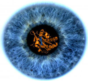 |
| Image of the iris of researcher Clemens Heikaus' eye with a model of a GAF domain imbedded in the pupil. A messenger molecule binds to the GAF domain to regulate an enzyme, PDE6, that is central to the way light hitting the retina is converted to signals to the brain. (Credit: Brad Clifton)
|
|
The section of the enzyme molecule that most interests the researchers is the so-called GAF A domain. A small messenger molecule, cGMP, binds to the GAF A domain to regulate the enzyme.
|
|
"The domain binds to this small molecule with extremely high sensitivity," said UW biochemist Clemens Heikaus, who along with Sergio E. Martinez, now a research associate at Rutgers, carried out the study. "From our structure, we can infer why it prefers cGMP over other messenger molecules." He added that the domain is quick in recognizing and responding to the messenger molecule to create an instantaneous flow of information to the brain.
|
|
Using X-ray crystallography and nuclear magnetic resonance, the researchers discovered that the enzyme undergoes major structural changes upon binding of the cGMP molecule.
|
|
Before binding occurs, the GAF domain is like an outstretched palm with the fingers wiggling, Heikaus said. After the cGMP molecule binds, the GAF domain closes and becomes less dynamic. In this state it looks more like a closed fist.
|
|
Further analysis of the consequences of this conformational change may lead to a better understanding of how the photoreceptor PDE helps regulate the path of signals that enable us to see, as well as provide general information on proteins with GAF domains.
|
|
"The addition of a simple, small molecule to the GAF domain affects the entire PDE enzyme," Heikaus said. Researchers think the binding to the domain may act as a switch that turns on the enzyme.
|
|
The research findings were published in the Sept. 19 Journal of Biological Chemistry. The article was selected as a Paper of the Week. The journal cover featured a striking image of the iris of Heikaus' eye, photographed by UW ophthalmology imaging supervisor Brad Clifton. Superimposed in the center of the pupil was a three-dimensional structure of the GAF domain.
|
|
In humans, GAF-containing proteins are rare. In plants and bacteria, GAF domains are widespread and are specialized for binding a variety of molecules. Some of these plant and bacteria GAF domains are important in detecting light, but they do so through a mechanism that is completely different from vision in vertebrate animals.
|
|
GAF domains emerged more than 3 billion years ago in early forms of life, and remained as animals and humans evolved, a phenomenon evolutionary biologists call conservation. Human GAF domains have similar protein folds, and a similar way of binding signal-triggering molecules inside a "pocket," as do GAF domains in more primitive creatures.
|
|
Humans have only a few kinds of GAF domains, all of which are in enzymes within the PDE family. They perform important functions not only in vision but also in hearts, lungs, and blood vessels. PDE5, an enzyme closely related to PDE6, is the therapeutic target for sildenafil, known by the trade name Viagra. In some men, this drug also inhibits PDE6 in the eyes, causing a temporary change in color vision.
|
|
More knowledge of the basic mechanisms of PDEs in vision may lead someday to better drug treatment for loss of eyesight from damaged retinas, such as occurs in night blindness and retinitis pigmentosa.
|