| Posted: October 11, 2008 |
International photo exhibitions includes nanotechnology images |
|
(Nanowerk News) Acclaimed scientists and photographers from around the world will share their scientific research, discoveries and observations of natural wonders in an international photography exhibition opening today at Rochester Institute of Technology.
|
|
RIT’s School of Photographic Arts and Sciences will host Images from Science 2 featuring 61 photographs from various scientific disciplines including astronomy, biology, engineering, medicine, oceanography, physics and nanotechnology. The opening, at 7 p.m. on Saturday, Oct. 11, takes place in SPAS Gallery, third floor of RIT’s Frank E. Gannett Building. The show runs through Oct. 25.
|
|
Viewers can see breathtaking images of the tiniest of things, from an unknown species of octopus measuring a half-inch in length to nanospheres with a diameter 300 times smaller than a human hair. Here are two examples:
|
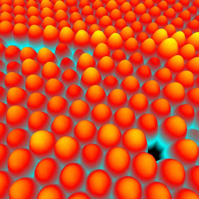 |
| Hans U. Danzebrink: Aesthetic Imperfections, 2008. Atomic force photomicrograph; magnification approximately x350,000 at capture; sample approximately 1.5 µm x 2.5 µm in size; post-capture colorization. Physikalisch-Technische Bundesanstalt (PTB), Braunschweig, Germany. This image reveals dislocations in a photonic crystal arrangement of polystyrene nanospheres magnified from a sample size of about 1.5 µm x 2.5 µm. In this case, the photonic crystals were grown as self-assembled structures from a colloidal solution. The nanospheres measure about 200 nm in diameter and are about 300 times smaller than a human hair, but still approximately 500 times larger than atoms. Practically, photonic crystals can be used to control and manipulate the flow of light, e.g. in optical waveguides. Even in nature, this type of nanosphere arrangement can be found. A classic example of this is observed in the gemstone opal with its iridescent play of light colors. Thorsten Dziomba of PTB provided the image data, and Dr. Frank Marlow of Max-Planck-Institut für Kohlenforschung supplied the sample. WSxM software was used to carry out the post-capture colorization.
|
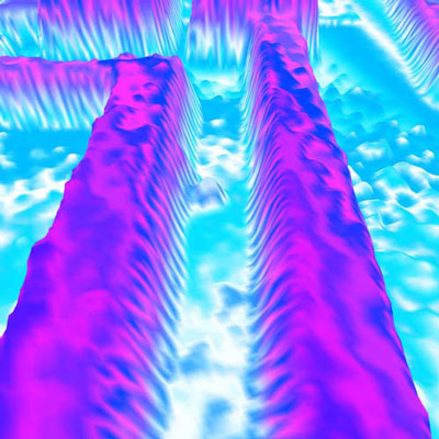 |
| Hans U. Danzebrink and Anna-Maria Gleixner. Data Channels, 2007. Atomic force photomicrograph; magnification approximately x35,000 at capture; actual sample approximately15 µm x 25 µm in size; post-capture colorization. Physikalisch-Technische Bundesanstalt (PTB), Braunschweig, Germany. This circuit-level image of a computer chip is a magnified view from a sample, which measures approximately 15 µm x 25 µm. The blue water-like coloring symbolizes the flow of information inside the microprocessor. The image data was provided by the nanometrologist Thorsten Dziomba, also of PTB, and the post-capture colorization was performed using WSxM scanning probe microscopy software for data visualization.
|
|
To accompany the display, RIT Cary Graphic Arts Press, the publishing arm of the Melbert B. Cary Jr. Graphic Arts Collection at RIT, produced a full-color companion catalog of all the images in the exhibition. The publication features an introduction by Martin Scott, a former director of scientific imaging at Eastman Kodak Co. The catalog will be available for purchase online through the RIT Cary Graphic Art Press at http://carypress.rit.edu and www.amazon.com.
|
|
“The quality of the images is excellent,” says Michael Peres, RIT department chair of biomedical photographic communications and one of the exhibit organizers. “All the traditional imaging methods are utilized, including micrography, high-speed nature photography, macro photography, but also some obscure methods such as scanning tunneling microscopy and radiography. It’s fascinating to see what people are currently doing in their respective scientific fields and the types of images they are producing.”
|
|
Scientists, photographers and worldwide institutions submitted entries for consideration. A photograph from Lennart Nilsson, a pioneer in medical photography, is among the images in the new exhibit. The award-winning Swedish photographer and scientist has been a leading supporter of the project since its inception. An international selection committee chose the 61 images from more than 300 entries. The final images were selected based on their scientific content, aesthetics and difficulty in making of the photograph.
|
|
In fall 2002, RIT launched the inaugural Images from Science exhibition. Since that time, Images from Science, has been hosted by 23 organizations in seven different countries, most recently in the Czech Republic.
|
|
“The first exhibition was so successful and far reaching because of the work produced by its outstanding contributors,” says Andrew Davidhazy, RIT department chair of imaging and photographic technology and one of the exhibit organizers. “Its longevity can be attributed to the stunning photographs that depict life as it is seldom seen by the general public. With this second exhibit, we wanted to once again emphasize to the photographic community that images made other than for artistic purposes can be appreciated not only for their scientific content, but also for their aesthetics.”
|
|
Adobe Systems Inc., Carl Zeiss MicroImaging Inc. and Durst Image Technology are sponsoring the project.
|
|
For more information, call RIT’s School of Photographic Arts and Sciences at (585) 475-2863 or visit the Images from Science Web site at http://www.rit.edu/cias/ritphoto/ifs-2008/photo_gallery/index.htm.
|