| Posted: October 21, 2008 |
German scientists advance light microscopy with contact-free laser technique |
|
(Nanowerk News) Although more than 400 years old, optical microscopy is still one of the most important methods used in medical research and biology. Its potential, however, could still increase significantly due to an invention recently made by a team of interdisciplinary working physicists.
|
|
Published as cover story in the current issue of the open access Journal Optics Express, the researchers from Prof. Käs' group in Leipzig, in collaboration with researchers in Jena and Cambridge present the Optical Cell Rotator (OCR), a novel tool for the contact-free orientation of biological samples using laser radiation ("The optical cell rotator").
|
|
According to first author Moritz Kreysing the new technology could be the missing key to allow for the realization of single-cell optical tomography, which has been shown in principle but not in any useful implementation.
|
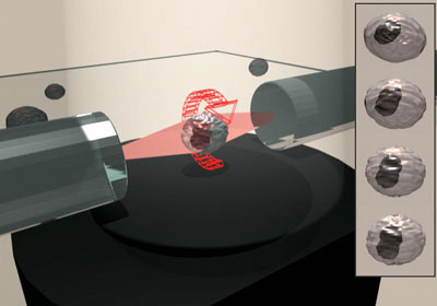 |
| A biological cell is held and rotated by laser beams emitted from two glas fibers.
|
|
In contrast to the current standards for 3D imaging of cells, namely confocal and deconvolution microscopy, tomographic imaging provides maximum resolution in all three dimensions. To achieve this feat specimens are rotated stepwise and imaged from multiple angles. Using this data a computer is able to reconstruct a three-dimensional model of the sample containing 2.5 times more information than traditionally accessible.
|
|
The Optical Cell Rotator is of fundamental importance in this context for the integrity of living samples. While the orientation of cells previously required their exposure to either strong electric fields or unphysiological mechanical tools, the new device promises a much more gentle handling of the sensitive material. Thus, OCR is probably most useful for the investigation of stem cells, which are notoriously sensitive to handling.
|