| Oct 07, 2013 |
Scientists create technique for high-speed, low-cost epigenomic mapping
|
|
(Nanowerk News) A new technique developed by researchers at the Stanford University School of Medicine could pave the way to an era of personalized epigenomics.
|
|
The technique, described in a study published online Oct. 6 in Nature Methods ("Transposition of native chromatin for fast and sensitive epigenomic profiling of open chromatin, DNA-binding proteins and nucleosome position"), could quickly yield huge amounts of useful information about which genes are active in particular cells. The technology involved is cheap, fast and easy to use, and all that would be needed from the patient is a blood sample or needle biopsy.
|
|
As word of the new technique has leaked, dozens of researchers around the world have begun putting it to work in their labs, said Howard Chang, MD, PhD, professor of dermatology at Stanford and a Howard Hughes Medical Institute early-career scientist. Chang shared senior authorship of the study with William Greenleaf, PhD, assistant professor of genetics. The lead author is graduate student Jason Buenrostro.
|
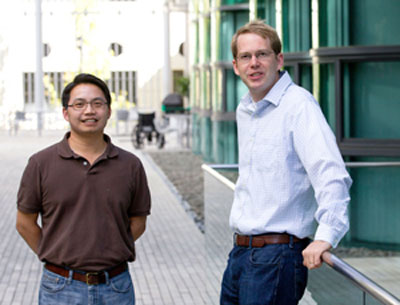 |
| The labs of Howard Chang, left, and William Greenleaf have developed a technique that could yield huge amounts of information about which genes are active in particular cells.
|
|
Genes are recipes for the production of proteins, which do almost all the work in every living cell. The biological field of genomics focuses on describing which genes an organism has. The newer field of epigenomics aims to discern which genes are actually used by various tissues within an organism — or, in the case of disease, misused. Virtually every cell in a person’s body contains essentially the same genes. Yet cells from different tissues — liver, skin, muscle, blood — do very different things because they use different genes, as do otherwise identical cells in different biochemical environments, developmental stages or states of health.
|
|
For a gene to give rise to the specific protein it codes for, the gene must be read and copied (or “transcribed”) by complex molecular machines. The genes of simpler, single-celled life forms, such as bacteria, are all available for transcription because those microbes’ DNA floats around as a flexible, circular chromosome within the cell.
|
|
But in complex organisms from yeast and amoebas to orchids and people, few of any cell’s genes are transcription-ready at any point in time. For example, humans’ much larger genomes feature vastly more DNA, much of which is devoted to regulating the timing and extent of each gene’s activation rather than encoding proteins. Human DNA, as well as that of other advanced life forms, is confined within a tiny cellular compartment known as the nucleus.
|
|
“If you could stitch together all 46 chromosomes in one of your cells and stretch the resulting, single string of DNA full-length, it would be about 2 meters long,” said Greenleaf. “But in real life, all that DNA is scrunched up inside the cell’s nucleus, which is about one two-hundred-thousandth of a meter in diameter.” That’s the rough equivalent, Greenleaf said, of bunching up a telephone line that stretches from New York City to Los Angeles and stuffing it into a two-bedroom house.
|
|
Most of a chromosome’s DNA is tightly spooled around ball-shaped protein assemblies called nucleosomes, rendering it inaccessible for transcription. Much of the genome is blocked this way. Elsewhere, various enzymes within the cell are capable of chemically relaxing some nucleosomes’ grip, unleashing erstwhile inaccessible DNA for transcription and hence altering the cell’s gene-use patterns.
|
|
Some of the current methods of determining the epigenomic state of a cell are so complex that only a handful of laboratories are equipped to carry them out. These procedures require dozens of separate technical steps, start-to-finish timescales of several days or more, and millions to tens of millions of cells from the same tissue.
|
|
In order to study relatively rare cell types using these methods, you have to do one of two things, said Chang. “You can force those cells to copy themselves repeatedly in the artificial environment of a laboratory dish or flask, driving their replication with biochemical sledgehammers. By the time you get enough cells for analysis, their epigenomic state may have changed wildly from its original condition.”
|
|
Alternatively, he said, you can pool biological samples from numerous different individuals. But this wipes out any possibility of meaningful personalized analyses.
|
|
The new method requires only 500 to 50,000 cells, which can easily be provided by one individual, Chang said. It involves about 15 minutes of hands-on technician time and takes as few as 10 hours from start to finish. Samples obtained on a daily basis from, say, a hospitalized patient or a subject in a clinical trial measuring a drug’s effect, can be processed in a clinically relevant time frame.
|
|
The insight that opened the door to this new technique came a year ago when the study’s lead author, Buenrostro, proposed that a bacterial enzyme could be used to “spray paint” the regions of the genome that are accessible to the molecular machines employed by cells to read genetic information.
|
|
Transposases — the kind of enzyme the Stanford scientists borrowed from bacteria — are found in all creatures. These enzymes insert copies of a particular DNA sequence into random sites along the genome. But because bacteria lack barriers such as nucleosomes, bacterial transposases haven’t evolved ways of inscribing their DNA “tags” on nucleosomally or otherwise blocked DNA.
|
|
The investigators used a bacterial transposase modified so that, instead of inserting its usual DNA tag at any part of the genome, it inserted special DNA sequences only in parts of the genome where nothing stood in the way. These DNA sequences were chosen to facilitate a high-throughput, laboratory-based, DNA-copying procedure. Incubating myriad copies of these sequences in a test tube, along with the modified transposase and a line of well-studied immune cells, yielded tags on whatever parts of the genome weren’t spooled around a nucleosome or occupied by one or another DNA-binding protein.
|
|
A single nucleosome ties up just under 150 chemical units of DNA. Tag-free DNA stretches of just that length designated nucleosome-blocked regions at numerous spots along the genome. Much shorter tag-free zones, numbering between eight and 10 DNA chemical units, occurred at specific sites along open areas of the genome known to be regulatory. These small, tag-free stretches, the Stanford team reasoned, had been occupied by DNA-binding proteins whose exact identities could be inferred from the size and sequence of the tag-free “footprint” they’d left. This was an important finding, as various DNA-binding proteins either facilitate or impede genes’ transcription.
|
|
To demonstrate the method’s clinical potential, the investigators drew blood from a healthy volunteer on three consecutive days, performing their analytic procedure each time. They were able to show, for this volunteer, which of three different regulatory DNA sites on a particular gene had been engaged by a DNA-binding protein. This regulatory-site pinpointing could guide clinical decisions about which drug would be best for changing a gene’s activity level with minimal side effects.
|