| Nov 04, 2013 |
Riboswitches in action
|
|
(Nanowerk News) A cell is a complex environment in which substances (metabolites) must maintain a correct state of equilibrium, which may vary depending on specific needs. Cells can maintain the proper concentrations of metabolites by regulating gene protein encoding through specific "switches", called riboswitches, which are able to block or activate protein synthesis. The precise mechanism by which these short strands of RNA carry out this function is still poorly understood. However, a study conducted by SISSA scientists Giovanni Bussi, Francesco Colizzi and Francesco De Palma and published in the journal RNA ("Ligand-induced stabilization of the aptamer terminal helix in the add adenine riboswitch"), now provides some important insights.
|
 |
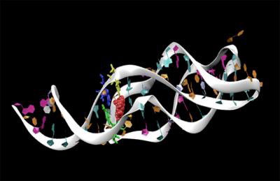
| This is an RNA strand.
|
|
A riboswitch is contained in a strand of messenger RNA, an RNA fragment that acts as a sort of template that "prints" the proteins that are needed for cell metabolism. However, unlike the rest of the messenger RNA unit, riboswitches don't actually encode any portion of the protein (i.e., they form part of what geneticists call non-coding RNA) but they serve to activate or deactivate the protein printing process and are located, in bacteria, in a stretch preceding the coding sequence. Scientists know that this switching action is made possible by a change in shape, which occurs when a part of the riboswitch (the aptamer) binds to a molecule in the cell environment which acts as a signal. Bussi and colleagues used computer simulations to reproduce the dynamics of the process and understand how binding to the metabolite brings about the change in shape.
|
|
More specifically, Bussi and colleagues simulated the riboswitch that uses an adenine molecule as a signal, to regulate the gene expressing a protein involved in the metabolism of adenine itself. Their findings clarify how adenine stabilizes the active form of the riboswitch (the one triggering protein synthesis) to the detriment of the inactive conformation. "We used molecular dynamics as a kind of 'virtual microscope' with which we observed the workings of the process", explained Bussi. "It's very important to understand these regulatory mechanisms since they are present in many bacteria – as well as in multicellular organisms – and may be useful for developing new antibiotics in the future."
|
|
The research was carried out within the framework of a European Research Council (ERC)-funded project coordinated by Bussi.
|
|
More in detail…
|
|
The computer-based simulations carried out in the study relied on PLUMED software, the latest release of which was presented in another recent paper published by Bussi in Computer Physics Communications. PLUMED is a software program devised to create and analyze simulations. "This more advanced version of the program was developed as a team of five young researchers from international institutions".
|
|
It's no coincidence that the researchers involved in this work are all young: the research project was in part funded by a special grant provided for young, non-permanent research fellows at SISSA and awarded to Bussi in 2011.
|

