| May 28, 2014 |
Ptychographic microscope for 3D live cell imaging
|
|
(Nanowerk News) Phase Focus Ltd (Phasefocus), the company that is revolutionising microscopy and imaging with the Phasefocus Virtual Lens®, reports on their latest publication released in Optics Express entitled "Ptychographic microscope for three-dimensional imaging".
|
|
Optics Express is the all-electronic journal for optics published by the Optical Society of America and edited by Andrew M. Weiner of Purdue University. It publishes peer-reviewed articles that emphasize scientific and technology innovations in all aspects of optics and photonics.
|
|
Label-free 3D live cell imaging is a challenge that remains to be addressed if advances in the development of 3D cell culture constructs for early-stage drug discovery are to reach their full potential. Improved label-free imaging methods are required that can simultaneously analyse all the cells within a 3D specimen rather than simply integrating a cumulative signal response.
|
|
In the latest paper based on ptychography, the authors from the universities of Sheffield & York together with UK company, Phasefocus, report on their latest research showing advances towards that goal. It is demonstrated that the technique may be used to visualise 3D specimens up to 34 tomographic sections in depth. These new results compare well with sectioned images collected from a confocal microscope but have the added advantage of strong phase contrast, which removes the need for sample labelling or staining.
|
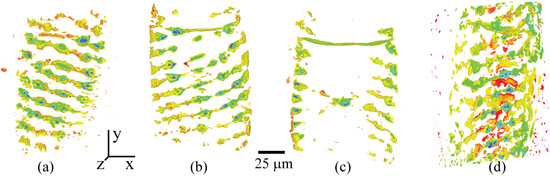 |
| The above images show the 3D volume rendering of a Spirogyra. Each image represents the total phase shift over 20 µm thick slices within the Spirogyra. (Images used by permission of T Godden of Phasefocus)
|
|
The technique is demonstrated on various samples including specimens of common Spirogyra algae and Volvox algae. In both cases, embedded animated images are available to the reader, one of the advantages of an online publication like Optics Express. The paper also reports images of Arabidopsis Thaliana plant embryo collected using ptychography together with those from a fluorescence confocal microscope; these comparisons clearly illustrate the excellent phase contrast that can be achieved with ptychography. The ptychography images are reconstructions of the optical phase shift throughout the whole specimen, whereas those from the confocal microscope represent the fluorescence intensity from the fluorophores which are embedded within the cell walls only.
|
|
In summary, the paper demonstrates how a form of 3D ptychography can be implemented on a conventional microscope platform and can produce optically sectioned images of relatively thick samples on the micron scale. It also demonstrates an increase in the number of sections, or slices, imaged using this method from 5 to 34. The Phasefocus 3D ptychographic method is unique in that it does not require rotation of the specimen with respect to the illumination source, or any translation of the specimen in the axial direction. This work clearly has potential applications in high contrast and stain-free live cell imaging, as well as in the imaging of thick, strongly scattering specimens using X-rays and electrons.
|
|
For more information on the Phasefocus VL21 live cell imaging system, please visit the Phasefocus web site: www.phasefocus.com/applications/cell-imaging/.
|