| Aug 11, 2014 |
Manipulating molecules to make memories
|
|
(Nanowerk News) Researchers have created a genetically engineered mouse that allows them to observe the movement of molecules in the brain that may be involved in the formation of memories. The transgenic mouse carries a fluorescently labeled messenger RNA (mRNA) that can be visualized in cells without disrupting normal physiological processes. This new genetic tool allows the observation of gene expression in real time in neurons where the movement of the fluorescently labeled beta-actin mRNA is providing clues to the molecular changes that take place in the brain to form and store memories.
|
|
The research team is based at Albert Einstein College, Bronx, NY, with collaborators in Novara Italy, Hunter College in New York, and the Howard Hughes Medical Institute in Ashburn, Virginia. They report their findings in Science ("Visualization of dynamics of single endogenous mRNA labeled in live mouse").
|
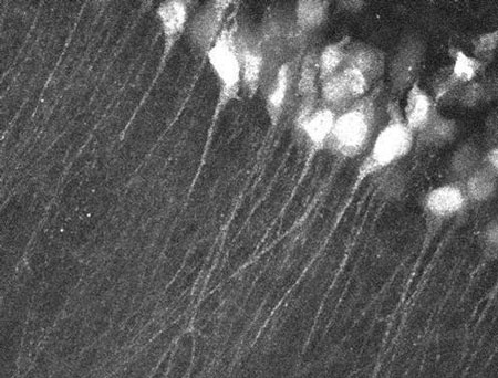 |
| Brain slice of the mouse hippocampus showing fluorescent beta-actin mRNA. (Image: Robert H. Singer, Einstein College of Medicine)
|
|
The group used a series of genetic manipulations to create a mouse whose natural beta-actin gene creates an mRNA that is fluorescently labeled and so can be observed in real time in the cells of the mouse. Beta-actin proteins are found in large amounts in numerous cell types, and are an important component of the structural scaffolding of the cell, which is involved in cell movement. However, the role of beta-actin in neurons has been less obvious. Study of brain slices from the transgenic mouse revealed that beta actin is transported to sites where two cells meet and transmit electrical and chemical signals to one another. These sites are known as cell synapses.
|
|
The study results demonstrate that the beta-actin protein is an essential structural protein in neurons that is important for strengthening neuronal contacts at the synapses, which are believed to play a key role in how memories are created and stored in the brain. The beta-actin works to strengthen those attachments to form what are known as dendritic spines. The strength of the attachment of the cells making up the dendritic spines appears to be a key factor in the establishment of a memory. Furthermore, the researchers identified a novel mechanism that can rapidly switch the beta-actin mRNAs from an active to an inactive state, which may be necessary for neuronal plasticity (the changing of neuronal shape and function) that may be involved in memory formation.
|
|
“These observations that neurons selectively activate protein synthesis and then shut it off fits very well with how we think memories are made,” said the senior author, Robert Singer, Ph.D., at Albert Einstein College of Medicine.
|
|
The investigators’ working hypothesis is that stimulation of the neuron results in controlled bursts of beta-actin protein synthesis, precisely where it is needed, to fortify the dendritic synapse and in doing so establish and strengthen the memory.
|
|
The observations reported here were of slices of brain from the transgenic mouse that were stimulated and observed in the laboratory. The investigators are now working on imaging technologies that would allow observation of the process in the intact brains of living mice. Those studies will seek supporting evidence for the current findings in addition to a more detailed understanding of the brain’s astonishing ability to manipulate molecules to make memories.
|