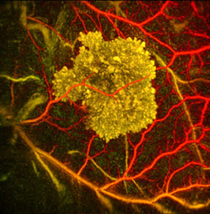
Photoacoustic image of Tyrosinase expressing cells. (Image: Paul Beard, Martin Pule)
The technique, developed by researchers at University College London with funding from the Biotechnology & Biological Sciences Research Council (BBSRC), combines a highly sensitive imaging system with a method of genetically engineering cells to produce a pigment which makes them more visible. The result is images of unprecedented clarity for organs at this depth within a live animal, without the need to inject a dye.
This opens up the possibility of studying cellular and genetic processes in mammals, as they happen.
Professor Martin Pule, University College London, one of the lead researchers on the study, said: "Anything you could possibly think of in terms of imaging complex activity within an organ, this technique would you allow you to do. Whether it's watching immune cells attack a tumour or an infection, or watching an organ develop embryonically, function, or react to damage or stress, all of these things you could observe at an organ level, which is something it would have been very difficult to do before.
"This technique lets you genetically label particular parts of an organ and then study it, over time, in a non-invasive way, without having to administer a contrast agent."
Other imaging methods either provide a highly detailed view of single cells within the uppermost millimetre of skin, or can penetrate deeper but only provide detail on the scale of a few millimetres. This device, based on a technique called photoacoustic imaging, allows scientists to distinguish features as small as clusters of tens of cells at depths close to a centimetre below the skin.
The photoacoustic imager contains a red laser, which shines pulses of light into the animal. Absorption of the light by pigmented cells produces sound waves which travel to the surface and are detected by an ultrasound scanner. By measuring the timings of the sound waves, the device builds up a picture of the cells.
Most photoacoustic scanners detect only blood vessels, as blood cells absorb most laser light. The UCL scientists have genetically engineered tumour cells so they create tyrosinase, the enzyme that produces the pigment melanin in skin. This turns the cells dark brown so they absorb light from the laser and can be detected by the photoacoustic device.
The researchers are now developing other pigments to increase the palette of colours available to label different parts of an organ, which would allow them to study complex behaviours of several different cell types.
Professor Melanie Welham, BBSRC Executive Director, Science, said: "Fundamental bioscience research is vital to reveal the biological mechanisms underlying normal physiology across the lifespan. Techniques such as this, which allow us to study the development and function of organs, have the potential to contribute to our understanding of human physiology and underpin efforts to improve health throughout the lifecourse."