| Jun 15, 2017 |
Novel application of CRISPR/Cas9 in plants - Visualizing DNA in living cells
|
|
(Nanowerk News) A research team around Andreas Houben from the Leibniz Institute of Plant Genetics and Crop Plant Research (IPK) in Gatersleben and Holger Puchta from the Botanical Institute of the Karlsruhe Institute of Technology developed a method to visualize defined genomic sequences in living plant cells and demonstrated its ability to reveal dynamic movements of chromosome ends (The Plant Journal, "Live cell CRISPR-imaging in plants reveals dynamic telomere movement").
|
|
This method allows the analysis of the spatio-temporal organization of the genome. It holds the potential to improve our understanding of how genome structure and function are intertwined.
|
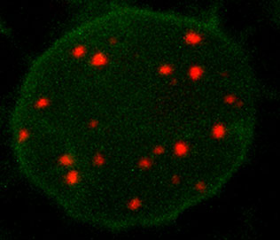 |
| Living nucleus of the plant Nicotiana benthamiana shows red fluorescent labelled telomere sequences. (Image: Steven Dreissig, IPK)
|
|
The spatial and temporal organization of genomes is important for maintaining and regulating cell functions such as gene expression, DNA replication and repair, and proper segregation of genetic material during cell division.
|
|
Therefore, elucidating how the genome is spatio-temporally organized inside the nucleus is imperative to understand how genes and non-coding DNA sequences are regulated during development. Mapping the functional organization of the genome can be achieved by visualizing interactions between different genomic elements in living cells.
|
|
“While fluorescence-tagged nuclear proteins can be readily imaged in living plant cells, in vivo visualization of defined DNA sequences turned out to be technically difficult”, explains Andreas Houben, head of the research group Chromosome Structure and Function of the IPK.
|
|
The discovery of the bacterial originated CRISPR-Cas9 system (type II clustered regularly interspaced short palindromic repeats CRISPR-associated caspase) revolutionized the field of the targeted genome editing in the last five years and has become a routine technology.
|
|
“By harnessing this system for live cell imaging in plants, our team shows the potential of this technology reaches far beyond the controlled induction of mutations”, explains Steven Dreissig, IPK scientist and first author of the study. “We demonstrate reliable imaging of telomere repeats located at the ends of the chromosome arms in living cells of Nicotiana benthamiana, a close Australian relative of tobacco, and pave the way for potential visualization of multiple genomic loci.”
|
|
“Furthermore, we show that CRISPR-Cas9 can be combined with fluorescence-labelled proteins to investigate DNA-protein interactions in living cells”, adds Holger Puchta, Director of the Botanical Institute in Karlsruhe.
|
|
This recent development may potentially enable plant scientists to visualize single genomic loci in living cells.
|