| Feb 06, 2013 |
Improved X-ray microscopic imaging
|
|
(Nanowerk News) X-ray microscopy requires radiation of extremely high quality. In order to obtain sharp images instrument and sample must stay absolutely immobile even at the nanometer scale during the recording. Researchers at the Technische Universitaet Muenchen and the Paul Scherrer Institute in Villigen, Switzerland, have now developed a method that relaxes these hard restrictions. Even fluctuations in the material can be visualized. The renowned journal Nature now reports on their results ("Reconstructing state mixtures from diffraction measurements").
|
|
For more than 100 years radiography meant: don't move! In order to visualize nanostructures such as biological cells, the porous structure of cement or storage fields of magnetic disks, the experimentators had to avoid any kind of vibration of X-ray microscope and sample. In addition, only a small percentage fraction of the incoming X-ray radiation could be used. Using special filters, they had to select exactly the fraction with the right properties – for example, the right wavelength.
|
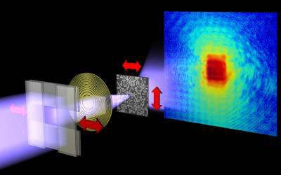 |
| While the test object is moved through the X-ray beam with nanometer precision, the scattered X-rays are captured by a detector. The scattering images are then reconstructed to an image of the sample.
|
|
Contributions of different wavelengths separated
|
|
Pierre Thibault of the Technische Universitaet Muenchen and Andreas Menzel, scientist at the Paul Scherrer Institute (Villigen, Switzerland) have now developed an interpretation method that produces reliable images in spite of vibrations or fluctuations. The method is based on a technique called "ptychography", developed in the 1960s for electron microscopy. Thibault and Menzel's advancements now make it possible to distinguish effects originating from the contribution of different types of X-ray waves.
|
|
Fluctuations visualized
|
|
Probably the most significant result of the study is that it gives access to a whole class of objects that previously could hardly be investigated. "We now not only can compensate for the vibrations in the microscope," says Andreas Menzel. "We can even characterize fluctuations of the sample itself, even if they are much too fast to be seen with individual snapshots."
|
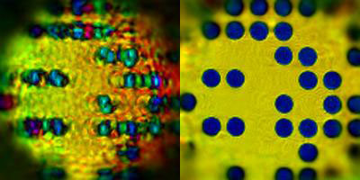 |
| Compared to a recording with conventional technology (left), ptychographic processing highly improves quality of the X-ray microscopic image (right).
|
|
"We needed to convince ourselves that the images we produced did indeed reflect accurately the samples and their dynamics," says Pierre Thibault. "So we carried out computer simulations. They confirmed that effects of the instrument as well as of the sample itself, such as flows, switching events or mixed quantum states, can be characterized."
|
|
Microscopic view inside
|
|
The new method combines the characterization of dynamical states with high-resolution X-ray microscopy. One possible application is to analyze the changing magnetization of individual bits in magnetic storage media with high storage density. The interactions of such single magnetic bits or their thermal fluctuations, which ultimately determine the lifetime of magnetic data storage, could be visualized.
|
|
"In addition to its use in imaging," explains Pierre Thibault, "our analysis method also reveals a fundamental relationship to other disciplines: Microscopy and scientific disciplines such as quantum computing, previously regarded as independent, can benefit from each other here."
|