| Apr 11, 2013 |
Synchrotron used as a microscope to determine protein structure
|
|
(Nanowerk News) A team led by David Reverter, a researcher at the Institute of Biotechnology and Biomedicine (IBB) of the UAB, has determined for the first time the three-dimensional structure of a protein pair: LC8 and Nek9. Depending on whether or not they bind, Nek9 ensures that the chromosomes group and separate correctly during cell division.
|
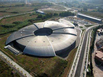 |
| This is the ALBA Synchrotron.
|
|
By analysing the 3D structure, these scientists have discovered a new mechanism that interferes with the protein binding and therefore also contributes to the correct regulation of cell division and other cell processes. The discovery could have implications for the study of diseases related to cell division processes, like cancer.
|
|
The 3D structure, published in the Journal of Biological Chemistry ("Structural analysis of the regulation of the DYNLL/LC8 binding to Nek9 by phosphorylation"), was obtained from data collected by the XALOC beamline of the ALBA Synchrotron. It is the first time that the crystal structure of a protein has been obtained by using this synchrotron and published in a scientific journal.
|
|
Taking part in the study were researchers from the Structural Biology Unit of the Institute of Biotechnology and Biomedicine (IBB) of the UAB, the Department of Biochemistry, Cell Biology and Molecular Biology of the University of Zaragoza, the Institute for Biocomputation and Physics of Complex Systems (BIFI), and the Institute for Research in Biomedicine (IRB Barcelona).
|