| Apr 18, 2013 |
Advanced nanofocusing optic enable studies of nanostructures with ultra-high resolution
|
|
(Nanowerk News) Microscopes have been a centerpiece of experimental science since at least the 16th century, providing a window into the material world at extraordinarily small scales. As the structures examined decrease in size – some measuring just billionths of a meter – capturing an x-ray image at high spatial resolution while retaining sufficient imaging contrast becomes more difficult.
|
|
One method of addressing this challenge is scanning x-ray microscopy, which uses a highly focused x-ray beam to produce spatial images of a specimen. In keeping with the scientific mission of Brookhaven National Laboratory's National Synchrotron Light Source II (NSLS-II), an advanced nanofocusing optic dubbed multilayer Laue lens (MLL) is being developed by the optics fabrication group for nanoscale imaging. The development has progressed to a prototype MLL microscope (see 2011 publication in Optics Express 19, 15069-15076).
|
|
For nanostructures that absorb x-rays very slightly, however, viewing them via absorption contrast becomes infeasible. Although the phase contrast provides a good way to study such a transparent object, historically researchers have found it difficult, if not impossible, to obtain accurate phase information of an object using x-rays because a detector only measures intensity. In scanning x-ray microscopy, previous attempts have been made to obtain quantitative phase information of a specimen, but none has worked well with MLLs, sacrificing the high-resolution power they provide for phase imaging.
|
|
As a result of years of groundbreaking research, a team of researchers in the Hard X-ray Nanoprobe (HXN) group at NSLS-II has solved this problem, together with their collaborators from Argonne National Laboratory, University of Connecticut and Chosun University in Korea. This research is detailed in a paper published in Nature's Scientific Reports, February 19, 2013 ("Quantitative x-ray phase imaging at the nanoscale by multilayer Laue lenses").
|
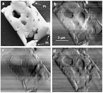 |
| (a) Scanning electron microscope (SEM) image of the solid oxide fuel cell (SOFC) specimen adhered on a Si3Ni4 window with Pt welding. (b-d) are horizontal phase-gradient scanning x-ray microscope images obtained by differential intensity, moment analysis and Fourier-shift fitting algorithms, respectively. Artifacts and blurring effects can be seen in (b) and (c), as compared to (d).
|
|
Brookhaven physicist Hanfei Yan, the paper's lead author, and his collaborators describe a new mathematical algorithm that clarifies, or "fits", the optical patterns that emerge from the scanning x-ray microscope, producing a quantitative phase image with far fewer artifacts and less blurring than existing methods. This novel technique pairs scanning x-ray microscopy with MLLs, fully exploiting its ultra-high resolution imaging capability for studies on nanostructures. When applied in a state-of-the-art light source like NSLS-II, a facility that will produce the brightest x-ray beams in the world, the nanofocusing power of MLLs will enable the study of nanostructures as small as 10 nm with an unprecedented level of precision.
|
|
The technique is so-called differential phase contrast imaging. Just as a beam of light "bends" when refracted by a prism, an x-ray beam can "bend" when refracted by a specimen. The characteristics of the specimen are then encoded in the emerging far field pattern, and by analyzing this pattern researchers are able to construct a quantitative phase image.
|
|
"Quantitative phase imaging at the nanoscale will enable us to examine the structural, compositional and possibly chemical state change of a specimen for which conventional contrast mechanism such as absorption and fluorescence imaging may not work," said Yan.
|
|
To verify the validity of this method, a solid oxide fuel cell (SOFC) anode sample composed of nickel and Yttria-stabilized zirconia cermet, a composite material made of ceramic and metallic elements, was investigated. When compared to images produced by conventional phase-imaging techniques, the superior image contrast of the method using an MLL microscope with a "fitting" algorithm is evident (see figure).
|
|
"The high sensitivity of the phase to structural and compositional variations makes this technique extremely powerful in correlating the electrode performance in SOFC," said Wilson Chu, a professor at University of Connecticut.
|
|
Currently, a next-generation MLL microscope with below 1-nm stability is under development, led by Evgeny Nazaretski, a co-author of the paper, and will be used as the workhorse at HXN beamline. "At NSLS-II, we will explore the full power of the MLL microscope and provide users not only a tool with high resolution, but also with new imaging capabilities," said Yong Chu, another co-author of the paper and also group leader of the HXN beamline.
|
|
Yan and his collaborators conducted the experiment at 26-ID of Advanced Photon Source at Argonne Lab, using a monochromatic x-ray beam with photon energy of 12 keV focused by a pair of multilayer Laue lenses placed orthogonally with respect to each other.
|