| Jul 19, 2013 |
A high-pressure nanoimaging breakthrough
|
|
(Nanowerk News) A team of researchers made a major breakthrough in measuring the structure of nanomaterials under extremely high pressures. Bragg coherent x-ray diffraction imaging (CXDI) is a promising tool to probe the internal strains of nanometer-sized crystals. But for high-pressure studies the x-ray beam must pass through a component of the diamond anvil cell, which can significantly affect the coherence properties of the beam. The researchers have developed a technique to deal with this that could lead to advances in new nanomaterials created under high pressures and a greater understanding of what is happening in planetary interiors ("Coherent diffraction imaging of nanoscale strain evolution in a single crystal under high pressure").
|
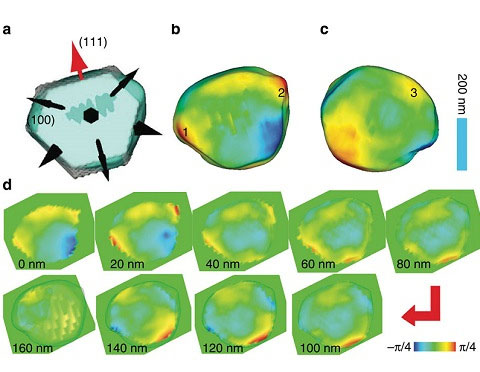 |
| (a) Isosurface (30%) of the reconstructed amplitude superimposed with a model of the possible {111} and {100} crystal planes. The normal directions of two sets of crystalline planes {111} and {100} are marked by two kinds of arrows (fat and narrow), and the one (111) used for the measurement is marked in red. (b,c) are the top and bottom view of phase shift distribution pasted on the 30% isosurface plot. Three strain distinguished locations numerically labelled are chosen for quantitative measurement as a function of pressure. (d) 3D phase distribution at different slicing depths spaced apart by 20 nm steps from top to bottom of the crystal. The colour scale is used to show the relative phase shift and normalized to range.
|
|
The team, with members from the Carnegie Institution of Washington, the Center for High Pressure Science and Technology Advanced Research (P.R. China), Argonne National Laboratory, University College London (England), and the Research Complex at Harwell (England) found that by averaging the diffraction patterns of the same crystal using different sample alignments in the instrumentation, and using an algorithm developed by researchers at the London Centre for Nanotechnology, they could compensate for the x-ray wavefront distortion passing through the pressure cell, and improve spatial resolution by two orders of magnitude.
|
|
The new technique is called the “mutual coherent function” method, or MCF. The researchers subjected a 400-nanometer (.000015-inch) single crystal of gold to pressures from about 8,000 times the pressure at sea level to 64,000 times that pressure, which is about the pressure in Earth’s upper mantle, the layer between the outer core and crust. The team conducted the imaging experiment at the X-ray Science Division 34-ID-C beamline at the U.S. Department of Energy Office of Science’s Advanced Photon Source at Argonne. They compressed the gold nanocrystal and studied it with CXDI, then applied the MCF method and found at first, as expected, that the edges of the crystal became sharp and strained. But to their surprise, the strains disappeared upon further compression. The crystal developed a more rounded shape at the highest pressure, implying an unusual plastic-like flow. Nanogold particles are very useful materials. They are about 60% stiffer compared with other micron–sized particles and could prove pivotal for constructing improved molecular electrodes, nanoscale coatings, and other advanced engineering materials.
|
|
The new technique will be critical for advances in these areas. Now that the distortion problem has been solved, the whole field of nanocrystal structures under pressure can be accessed and the scientific mystery of why nanocrystals under pressure are somehow up to 60% stronger than bulk material may soon be unraveled.
|