| Nov 06, 2013 |
Nanoparticles reveal mechanisms of cancer cell growth in whole cells
|
|
(Nanowerk News) Healthy cells are renewed by dividing and dying off, but cell division in cancer cells goes unchecked because natural cell death is suspended. This happens because too many receptors for the growth factor EGF which are found on the surface of the cell join together to form pairs. These pairs start a signal chain into the cell, culminating in unrestricted growth. Now, scientists at the INM – Leibniz Institute for New Materials have for the first time been able to show this pairing in human cancer cells on individual receptors using gold nanoparticles.
|
|
The results were recently published in the online journal Scientific Reports ("Epidermal growth factor receptor subunit locations determined in hydrated cells with environmental scanning electron microscopy").
|
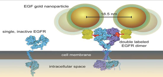 |
| Labeling of the EGFR with gold nanoparticles vizualizes its dimerisation. (Source: INM)
|
|
“It has never before been possible to show the mechanism of pairing in individual receptors in whole cells”, explains Diana Peckys, a human biologist at the INM. “Up to now, biochemical methods in which the cells are either in principle destroyed or only ever receive calculated mean values from the observation of many receptors have been used”, she goes on. “We examined the arrangements of individual receptors in pairs and in smaller groups. This was possible because we were able to show the individual receptors on the intact cell under an electron microscope.”
|
|
To do this, researchers marked the growth factor EGF using gold nanoparticles with a diameter of around ten nanometers. At the same time, they worked with a special measuring and preparation technique that makes it possible to examine whole cells in their natural fluid state in the nanometer range under a scanning electron microscope. They used these combined methods with a resolution of three nanometers to for the first time show on a one to one basis that EGF binds to the receptor and form pairs and clusters.
|
|
In addition to experimentally proving previous theoretical calculations, the team from the Innovative Electron Microscopy Program Area is now stepping up its work with German cancer researchers. “With our new measuring method, we are keen to focus in the future on investigating how different cancer drugs influence pairing and grouping of the EGFR and similar related receptors. Observing these processes in terms of individual molecules on intact cells opens up new prospects for cancer research”, the electron microscope expert sums up.
|
|
Background
|
|
All cells in mammalian organisms have receptors for the epidermal growth factor (EGFR). If the related growth factor (EGF - epidermal growth factor) combines with the EGFR receptor, the receptor changes shape so that it can join together with a second receptor to which a growth factor can then also readily bind. Through a complicated signal chain, an activated receptor pair such as this promotes the cell growth that in healthy cells produces normal cell renewal, but if too many of these so-called dimers are formed on the surface of the cell , this stimulates the cells to produce excessive cell division and growth, and this can lead to malignant tumours and metastases.
|