| Posted: Jul 09, 2009 | |
Nerve interface electrodes |
|
| (Nanowerk Spotlight) The potential of micro- and especially nanotechnology applications in neuroscience is widely accepted. Different biomedical devices implanted in the central nervous system, so-called neural interfaces, already have been developed to control motor disorders or to translate willful brain processes into specific actions by the control of external devices. These implants (see also "Brain implants improved by nanotechnology coatings") could help increase the independence of people with disabilities by allowing them to control various devices with their thoughts. | |
| A recent example of this emerging brain technology is an electrode for a peripheral nerve interface developed at AIST and Toyohashi University of Technology in Japan. | |
| Neural interfaces used for such purposes as electroencephalography are noninvasive, but suffer from relatively poor spatial and temporal resolution of signals. The type of neural interface that uses electrodes inserted in the brain and measures neuronal activities is more effective, but might leave behind irreversible lesions in the cerebrum because of the need to implant electrodes in brain tissue. Other problems with this type of neural interface include the difficulty of obtaining information about individual organs. | |
| Believing that an effective solution to these problems lies in designing a neural interface that attaches not to the cerebrum but to peripheral nerves, Hidekazu Kaneko from the Institute for Human Science and Biomedical Engineering at AIST and his team have been working with Makoto Ishida's Integrated Circuit Group at Toyohashi University of Technology to develop an electrode for a peripheral nerve interface. | |
| According to Kaneko, the electrode under development should have the capability of simultaneously measuring the action potentials of individual nerve fibers in a peripheral nerve bundle. Earlier proposals for measuring peripheral nerve activity were based on the use of sieve electrodes, needle point holder-shaped electrodes, and cuff electrodes; but these and other attempts have been unable to meet the requirement for low invasive measurement that is also able to distinguish the activity of individual nerve fibers. | |
 |
|
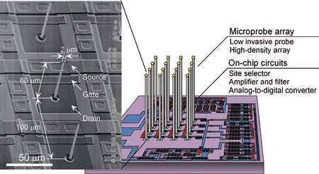
| Fig. 1: Microprobe electrode array fabricated by VLS growth. This is an electron microscope photograph of the microprobe electrode array under development (left). Conventional microelectrodes are thick, typically from several tens to a hundred micrometers in diameter. The microprobes shown here are extremely fine with a diameter of only 2 micrometers, for low-invasive use. Moreover, these microprobes can be grown on a semiconductor substrate (right). (Photo and illustration provided by Makoto Ishida, Toyohashi University of Technology) | |
| Applying the selective Vapor-Liquid-Solid (VLS) growth technique to electrode development, the team has succeeded in forming a structure like that shown in the electron microscope photograph (Fig. 1, left). | |
| The resulting electrode combines an unprecedented low-invasive design with ease of incorporation on an integrated circuit substrate. Moreover, using an array of metal microelectrodes having similar recording surface area as this electrode, the researchers have confirmed the ability to take localized measurements of evoked action potentials in single peripheral nerve fibers (Fig. 2) for all-or-nothing responses. | |
 |
|
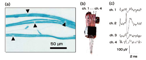
| Fig. 2: Measurement of action potentials in a single peripheral nerve fiber. On myelinated peripheral nerve fibers, action potentials occur at the Ranvier nodes (the triangles in tissue sample (a)). Using a microelectrode array (b) consisting of metal microelectrodes arranged at approximately 0.5 mm intervals, we were able to take localized measurements of evoked action potentials in single nerve fibers (c) for all-or-nothing responses. | |
| The measured signals attenuated with increased distance from the signal source, showing that this new technology for simultaneous measurement of action potentials in multiple neurons is able to isolate action potentials for individual nerve fibers. | |
| In the future, the team hopes to enable use of this peripheral nerve electrode for measuring nerve activities and for controlling them by electrical stimulation, so that the technology can be applied to assisting and restoring organs whose functionality is impaired due to diseases and other causes. | |
| From an article by Hidekazu Kaneko in AIST Today | |
|
Become a Spotlight guest author! Join our large and growing group of guest contributors. Have you just published a scientific paper or have other exciting developments to share with the nanotechnology community? Here is how to publish on nanowerk.com. |
|