| Posted: Sep 11, 2009 | |
Nanoparticles' random walk has implications for nanotoxicology |
|
| (Nanowerk Spotlight) The interest in exploring the use of noble metal nanoparticles – such as gold, silver and their alloys – for diagnostic and therapeutic imaging stems from the drawbacks of current in vivo probes. Fluorescent probes, such as fluorescent dyes and proteins, are not photostable and therefore are useful only for a limited time during the probing event. Besides imaging agents, especially gold nanoparticles are also intensely researched as target-specific vehicles for drug delivery. | |
| Due to its inert chemical properties, gold has been widely considered as one of the most stable and biocompatible materials. But, as X. Nancy Xu explains to Nanowerk, "studies of the biocompatibility and toxicity of gold nanoparticles in various types of cells, have yielded inconclusive results: some studies show a toxic effect and high-dependence of toxicity on nanoparticle size and surface functional groups, while other studies report no significant cytotoxicity. Many of these studies did not use purified gold nanoparticles, or examine any other chemicals present in the gold nanoparticle solutions, or well characterize the physical properties (e.g., possible size change and aggregation) of gold nanoparticles in buffer solution and cell culture media during the experiments, leading to these inconclusive results. Furthermore, study of the biocompatibility and toxicity of gold nanoparticles in living animals is yet to be fully explored." | |
| Xu is a professor in chemistry and biochemistry at Old Dominion University in Norfolk, Virginia. In her latest work, she and her group have synthesized and characterized stable (photostable, non-aggregating), nearly monodisperse, and highly purified gold nanoparticles, and utilized them to study cleavage-stage embryos in real-time and to probe their effects on embryonic development at the single-nanoparticle level in real time. | |
| The team has reported their findings in a recent paper in Nanoscale ("Random walk of single gold nanoparticles in zebrafish embryos leading to stochastic toxic effects on embryonic developments" – free access article). | |
 |
|
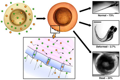
| Schematic of the research findings. Gold nanoparticles accumulate in the embryos. Optical images of zebrafish at a given concentration of gold nanoparticles. (Image: Lauren M. Browning, X. Nancy Xu, Old Dominion University) | |
| "We found that single gold nanoparticles passively diffused into chorionic space of the embryos via their chorionic pores and continued their random-walk into inner mass of embryos," says Xu. "Diffusion coefficients of single nanoparticles vary dramatically as nanoparticles diffuse through various parts of embryos, suggesting highly diverse transport barriers and viscosity gradients of the embryos." | |
| The researchers also found that the amount of gold nanoparticles accumulated in embryos increases with increased nanoparticle concentration. | |
| "Interestingly" says Xu, "their effects on embryonic development show random dependence on concentration – they are not proportionally related to their concentration." | |
| The team found that the majority of embryos (74% on average) chronically incubated with 0.025–1.2 nM gold nanoparticles for 120 hours developed to normal zebrafish, with some (24%) being dead and a few (2%) deformed. This result is in stark contrast with what they reported previously for silver nanoparticles (we have covered this in a previous Nanowerk Spotlight about a real-time study of nanosilver in fish embryos), showing that gold nanoparticles are much more biocompatible with the embryos than silver nanoparticles. This also suggests that the biocompatibility and toxicity of nanoparticles depends on their chemical properties. | |
| It is well known that the physical and chemical properties of nanoparticles are highly dependent upon a range of parameters – their size, shape, surface properties, embedded solvents, and the way that they were prepared and purified. | |
| As Xu points out, their chemical and physical properties will surely affect their interactions with living organisms, and define their biocompatibility and toxicity in given living organisms. "Therefore, it will be misleading if one tries to compare the study of one type of nanoparticles in one living organism with other types of nanoparticles in other living organisms" she says. | |
| To overcome the limitations of current nanotoxicity studies, Xu's team have developed three important components for such studies: | |
| – new methods to prepare stable (non-aggregated) and purified model nanoparticles (e.g., different sizes and surface functional groups of gold and silver nanoparticles); | |
| – real-time imaging tools (e.g., DFOMS) for characterizing the size of individual nanoparticles in vivo in real-time; and | |
| – effective in vivo assays (zebrafish embryos) for screening and probing the biocompatibility and toxicity of model nanoparticles, aiming to depict the dependence of biocompatibility and toxicity of nanoparticles on their physical and chemical properties, and their underlying mechanisms. | |
| "We found gold nanoparticles in various parts of normally developed zebrafish," says Xu. "Together with the strong variations in diffusion coefficients, these interesting findings suggest that the random diffusion of gold nanoparticles in embryos during their development might have led to uncertain effects on embryonic development." | |
| The team is already working on further probing what causes the embryos to become normally developed, deformed or dead zebrafish, as they are incubated with nanoparticles. | |
 By
Michael
Berger
– Michael is author of three books by the Royal Society of Chemistry:
Nano-Society: Pushing the Boundaries of Technology,
Nanotechnology: The Future is Tiny, and
Nanoengineering: The Skills and Tools Making Technology Invisible
Copyright ©
Nanowerk LLC
By
Michael
Berger
– Michael is author of three books by the Royal Society of Chemistry:
Nano-Society: Pushing the Boundaries of Technology,
Nanotechnology: The Future is Tiny, and
Nanoengineering: The Skills and Tools Making Technology Invisible
Copyright ©
Nanowerk LLC
|
|
|
Become a Spotlight guest author! Join our large and growing group of guest contributors. Have you just published a scientific paper or have other exciting developments to share with the nanotechnology community? Here is how to publish on nanowerk.com. |
|