| Posted: Sep 22, 2009 | |
Cell surface engineering with DNA nanotechnology |
|
| (Nanowerk Spotlight) Bionanotechnology researchers are experimenting with techniques for attaching DNA nanoarrays to cell surfaces for various reasons: to label cell surfaces with functionalized micrometer-sized patches; to deliver materials such as nanoparticles or carbon nanotubes to cell surfaces; to deliver nucleic acids into the cell for gene silencing; or to engineer microtissues of cell/cell networks by using self-hybridizing properties of single stranded DNA molecules. | |
| A team of scientists in California has now successfully attached self-assembled DNA structures to cancer cells using two different methods. This is one of the first illustrations of the biomedical relevance of DNA arrays and but one example where nanotechnology is used today. | |
| "We have demonstrated how self-assembled DNA arrays can be directed to the surface of cells, first through biotin-streptavidin interactions and second through specific antibody-cell surface interactions," Alexey Koyfman tells Nanowerk. "We also explored the versatile cargo-carrying ability of these DNA arrays for directing cell-surface interactions, cell-cell bridging, and positioning multiple cells onto a DNA fabric. Our assembly strategy is simple and effective, although the preparation of more monodisperse arrays as well as control of the organization, density, and purification of specific biomolecule couplings is required for more complicated applications." | |
 |
|
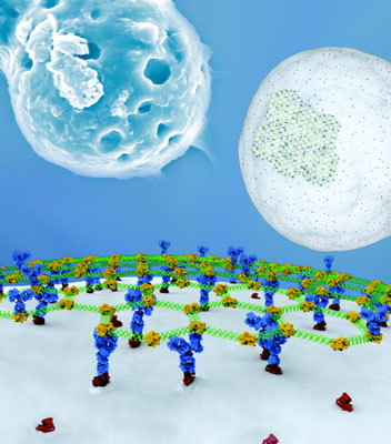
| Self-assembled DNA arrays can be directed to the surface of cells through specific antibody-cell surface interactions. The versatile cargo-carrying ability of arrays is explored for directing cell-surface interactions, cell-cell bridging, and positioning multiple cells onto a DNA fabric. (Illustrated by Peter Allen, UCSB) | |
| Koyfman was a graduate student in Norbert Reich's Biochemistry Lab at UC Santa Barbara when this work was done and is now a postdoctoral fellow at the National Center for Macromolecular Imaging at Baylor College of Medicine. Back in January we reported about his work on assembling three-dimensional DNA nanostructures ("DNA nanotechnology goes 3D "). | |
| The new results, also first-authored by Koyfman, have been reported in the September 16, 2009 online issue of Journal of the American Chemical Society ("Cell-Targeted Self-Assembled DNA Nanostructures") | |
| Most cell surface engineering is done by chemically modifying surfaces of cell. Reich's group, however, are modifying cell surfaces by attaching DNA cell patches. With their technique, they can also build microtissues by enclosing cells together using large DNA arrays. | |
| Koyfman says that it is also possible that the fate of stem cells could be induced using their targeted DNA architectures carrying molecular cargo since previous research showed that stem cell fate could be controlled by external small molecules. | |
| The team assembled their arrays from three single-stranded DNA molecules into hexagonal units repeated every 30 nanometers and into extended networks spanning several micrometers. Their first strategy for attaching DNA arrays to cells involves a nonspecific route using lysine-reactive biotin. Their second strategy for directing DNA arrays to cancer cells builds on the previous strategy but uses interactions between cell-specific surface proteins and their antibodies. | |
| There are three potential applications of this DNA nanotechnology work. Larger DNA arrays, that are hundreds of microns long, could be used to encapsulate a number of cells resulting in cell-cell networks that may be useful in tissue engineering. Smaller DNA arrays could be used for molecular delivery to cell surface, or, binding to single cells and coupled to fluorescent molecules, could be used for cellular screening. | |
 |
|
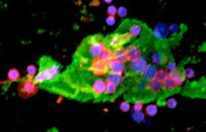
| Confocal microscopy of a large DNA-biotin-STV-Ab array bound to epidermal growth factor receptors on multiple HeLa cells. DNA arrays are green and HeLa cell surfaces red; HeLa cytoplasm is blue (Image: Dr. Koyfman). | |
| Koyfman says that one of the challenges is controlling the DNA scaffold size bound to a cell. "By controlling the DNA patch size, different applications are possible. The large and small sizes of DNA arrays need to be optimized before we can determine which application is the most promising." | |
 By
Michael
Berger
– Michael is author of three books by the Royal Society of Chemistry:
Nano-Society: Pushing the Boundaries of Technology,
Nanotechnology: The Future is Tiny, and
Nanoengineering: The Skills and Tools Making Technology Invisible
Copyright ©
Nanowerk LLC
By
Michael
Berger
– Michael is author of three books by the Royal Society of Chemistry:
Nano-Society: Pushing the Boundaries of Technology,
Nanotechnology: The Future is Tiny, and
Nanoengineering: The Skills and Tools Making Technology Invisible
Copyright ©
Nanowerk LLC
|
|
|
Become a Spotlight guest author! Join our large and growing group of guest contributors. Have you just published a scientific paper or have other exciting developments to share with the nanotechnology community? Here is how to publish on nanowerk.com. |
|