| Posted: Feb 01, 2007 | |
One day doctors will grow new bones with nanotechnology |
|
| (Nanowerk Spotlight) Nanotechnology-enabled tissue engineering is receiving increasing attention. The ultimate goal of tissue engineering as a medical treatment concept is to replace or restore the anatomic structure and function of damaged, injured, or missing tissue. | |
| At the core of tissue engineering is the construction of three-dimensional scaffolds out of biomaterials to provide mechanical support and guide cell growth into new tissues or organs. Biomaterials can be variously permanent or biodegradable, naturally occurring or synthetic, but inevitably need to be biocompatible. Using nanotechnology, biomaterial scaffolds can be manipulated at atomic, molecular, and macromolecular levels. | |
| Creating tissue engineering scaffolds in nanoscale also may bring unpredictable new properties to the material, such as mechanical (stronger), physical (lighter and more porous) or chemical reactivity (more active or less corrosive), which are unavailable at micro- or macroscales. For bone tissue engineering, a special subset of osteoinductive, osteoconductive, integrative and mechanically compatible materials are required. Such materials need to provide cell anchorage sites, mechanical stability, structural guidance and an in vivo milieu. | |
| Moreover, they need to provide an interface able to respond to local physiological and biological changes and to remodel the extracellular matrix (ECM) in order to integrate with the surrounding native tissue. Scientists in Singapore have developed a new nanoscale biocomposite that brings researchers one step closer to mimicking the architecture of the ECM. | |
| Nanofibers and nanocomposites are highly promising recent additions to materials in relation to tissue engineering. To achieve the goal of tissue reconstruction, nanofibrous scaffolds must meet some specific requirements: A high porosity and an adequate pore size are necessary to facilitate cell seeding and diffusion throughout the whole structure of both cells and nutrients. Biodegradability is essential since scaffolds need to be absorbed by the surrounding tissues without the necessity of a surgical removal. The rate at which degradation occurs has to coincide as much as possible with the rate of tissue formation. | |
| The researchers in Singapore, together with colleagues from the University of London and the Indian Institute of Technology, used an operationally simple electrospinning technique to fabricate polycaprolactone/nanohydroxyapatite/collagen (PCL/nHA/Col) biocomposite nanofibrous scaffolds to provide mechanical support and to direct the growth of human fetal osteoblasts (hFOB). | |
| "While cells are fabricating their own natural matrix structure around themselves, our newly designed nanofibrous scaffold is able to provide structural integrity within the body and eventually it will break down leaving the neotissue, newly formed tissue which will take over the mechanical load for the repair of diseased tissues or organs" Jayarama Venugopal explains to Nanowerk. "We found that our unique nanoscale biocomposite system had inherent surface functionalization for hFOB adhesion, migration, proliferation and mineralization to form a bone tissue for the regeneration of bone defects." | |
| Venugopal, a researcher at the Nanoscience and Nanotechnology Initiative, Division of Bioengineering, at the National University of Singapore, is first author of a recent paper published in Nanotechnology ("Biocomposite nanofibers and osteoblasts for bone tissue engineering"). | |
  |
|
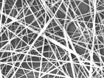
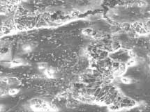
| Left: PCL/nHA/Col biocomposite nanofibers (373 ± 191 nm); Right: Multilayer of hFOB cells on PCL/nHA/Col biocomposite nanofibers (Reprinted with permission from IOP Publishing) | |
| "The resulting nanofibrous scaffolds were highly porous (>80%) structures and provided a sufficient open pore structure for cell occupancy whilst allowing free transport of nutrients and metabolic waste products" says Venugopal. "Moreover, vascular in-growth was facilitated. The mineralization was significantly increased (55%) after 10 days of culture and appeared as minerals synthesized by osteoblast cells." | |
| Surface topography of nanostructured substrate plays a critical role in regulating initial cell behavior, such as cell adhesion, which can also influence cellular viability and proliferation. Venugopal points out that the fibrous architecture of their biocomposite material mimicked the natural ECM and assisted in maintaining a normal phenotype of the cells. | |
| "Our results also suggest that there may be advantages for osteoblast cells to be cultured in vitro on nanofibrous scaffolds resembling the architecture of bone "Venugopal notes. "This could be of clinical significance for the use of tissue engineering in repairing of complicated bone defects." | |
| Nanotechnology for tissue engineering applications is just started, and most of the applications are still in the early research stage. Research like this one will ultimately lead to the fabrication of nanofibrous scaffolds for dermal wound healing (wound cover), bone filling, skin, bone, cartilage, nerve, and vascular tissue engineering. | |
 By
Michael
Berger
– Michael is author of three books by the Royal Society of Chemistry:
Nano-Society: Pushing the Boundaries of Technology,
Nanotechnology: The Future is Tiny, and
Nanoengineering: The Skills and Tools Making Technology Invisible
Copyright ©
Nanowerk LLC
By
Michael
Berger
– Michael is author of three books by the Royal Society of Chemistry:
Nano-Society: Pushing the Boundaries of Technology,
Nanotechnology: The Future is Tiny, and
Nanoengineering: The Skills and Tools Making Technology Invisible
Copyright ©
Nanowerk LLC
|
Become a Spotlight guest author! Join our large and growing group of guest contributors. Have you just published a scientific paper or have other exciting developments to share with the nanotechnology community? Here is how to publish on nanowerk.com.