| Posted: Jan 18, 2010 | |
New modality of force microscopy advances non-destructive subsurface characterization techniques |
|
| (Nanowerk Spotlight) In characterizing materials, especially live biological specimen such as cells, it is important not only to be able to explore the surface but also any subsurface structures and properties – without damaging or destroying the sample and for hard and soft materials alike. For example, many synthesized nanoparticles can readily get inside a cell. Therefore studying the cell surface, while useful, can provide little or no knowledge about the particles hidden in the interior of the cell. Another example is the detection of nanoscale defects in nanofabricated structures such as those made by electron beam lithography; or the detection of embedded cracks and voids in nanocomposite materials. | |
| A promising technology developed in the early 1990s is ultrasonic force microscopy. In one variation of this technique, a high-frequency acoustic wave – with a much higher frequency than that of a typical scanning probe cantilever – is launched from the bottom of the specimen while another wave with a slightly different frequency is launched from the cantilever. The interference of these two waves would nominally form a surface acoustic standing wave. Perturbations to the phase and amplitude of the surface acoustic standing wave are locally monitored by the scanning probe microscope. As the specimen acoustic wave is perturbed by buried features, the resultant alteration in the surface acoustic standing wave, especially its phase, is effectively monitored by the SPM cantilever. Scanning a sample can then be translated into a visual representation of its acoustic wave perturbation which offers a quantitative account of the internal features of the specimen (for more details see "Imaging nanoparticles in cells by nanomechanical holography" and "Elastic phase response of silica nanoparticles buried in soft matter"). | |
| "We have shown that an atomic force microscope can obtain a range of surface and subsurface information by making use of the nonlinear nanomechanical coupling between the probe and the sample," Ali Passian, a researcher with the Nanoscale Science and Devices Group at the Oak Ridge National Laboratory (ORNL), tells Nanowerk. "This technique, which we call mode-synthesizing atomic force microscopy (MSAFM), relies on multi-harmonic forcing of the sample and the probe." | |
| Passian explains that MSAFM, using nonlinear interactions, in a single run is capable of delivering myriad nanoscale features not attainable by other means. The probe is a micrometer sized solid oscillator, generally made of silicon, which is brought very close to the material under study. By carefully controlling the dynamics of the interacting probe-sample, one can obtain a host of surface and subsurface material properties. | |
| "Our work also shines a new light on the way to look at the different techniques using mechanical excitation of the probe or the sample – tapping mode, ultrasonic force microscopy, bimodal, etc." says Passian. The technique is flexible to accommodate and describe all these modes and provides a clearer understanding of the richness of the (mechanical) information available to characterize materials at the nanoscale." | |
 |
|
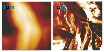
| MSAFM image of a poplar cell wall in the configuration of three excitation states.(left) Standard AFM topography image (7µm scan size) of a poplar cell wall. (right) MSAFM image of the same region. (Reprinted with permission from Nature Publishing Group) | |
| Passian and his collaborators from ORNL, Laurene Tetard and Thomas Thundat, have published their work in the December 20, 2009 online edition of Nature Nanotechnology ("New modes for subsurface atomic force microscopy through nanomechanical coupling"). | |
| The team has demonstrated the versatility of their methodology by showing the controlled use of the synthesized modes for surface and subsurface characterization of a structurally simple nanofabricated sample and poplar cells. | |
| "Our findings suggest a potential application in studying complex samples such as an organic system that shows a variety of interrelated chemical, morphological and mechanical properties, as opposed to simple samples characterized rather with homogeneity, uniformity and isotropicity," Passian points out. | |
| In previous applications of ultrasonic microscopy, the probe-sample interaction – which is manifested by the nonlinear force experienced by the very sensitive probe – was not fully utilized and therefore the acquired information was limited. | |
| "In nonlinear optics wave mixing can occur in a material with a nonlinear polarizability which results in a set of new frequencies that are currently used in a number of important applications," explains Passian. "In an analogues manner, using the nonlinear tip-sample interaction in place of a nonlinear medium, the mixing of ultrasonic waves can now be exploited to a higher level of coupling than the simple difference between the two excitations." | |
| Since MSAFM can help the acquisition of a host of both topographic as well as subcellular information in a very sensitive manner, the ORNL team suggests that it could not only be used as a dynamic sensor but also to detect vibrations/natural resonance frequencies of a multi-component systems such as a living cell or structures with complex molecules. | |
 By
Michael
Berger
– Michael is author of three books by the Royal Society of Chemistry:
Nano-Society: Pushing the Boundaries of Technology,
Nanotechnology: The Future is Tiny, and
Nanoengineering: The Skills and Tools Making Technology Invisible
Copyright ©
Nanowerk LLC
By
Michael
Berger
– Michael is author of three books by the Royal Society of Chemistry:
Nano-Society: Pushing the Boundaries of Technology,
Nanotechnology: The Future is Tiny, and
Nanoengineering: The Skills and Tools Making Technology Invisible
Copyright ©
Nanowerk LLC
|
|
|
Become a Spotlight guest author! Join our large and growing group of guest contributors. Have you just published a scientific paper or have other exciting developments to share with the nanotechnology community? Here is how to publish on nanowerk.com. |
|