| Posted: Mar 16, 2007 | |
Nanotube biological probes for intracellular studies |
|
| (Nanowerk Spotlight) The assembly of nanoparticles along the external or internal surface of carbon nanotubes (CNTs) is of both fundamental and technological interest. Combining unique properties of CNTs and nanoparticles, the nanoparticle/nanotube composite structure attracts a broad range of advanced applications, including nanoelectronics, chemical and biosensors, catalysis and fuel cells. | |
| This so-called 'decoration' of CNTs has been used to increase the hydrogen storage capacity, to make nanotubes magnetic, or to grow secondary structures inside the nanotubes to increase the available surface for catalysis. In the case of interior wall decoration of CNTs, the internal cavity of the nanotube often is obstructed and no flow can be achieved or there could be release of the particles in the environment. | |
| In the case of exterior wall decorations, the particles enter in direct contact with the environment and may be lost during the nanotube handling. A novel technique of multifunctional nanotubes with controllable amounts of nanoparticles embedded in their walls during the synthesis process solves both problems leaving the CNT bore accessible and keeping the nanoparticles shielded from the environment by the CNT walls. This paves the way to using carbon nanotubes as nanoscale biological probes for sub-cellular investigation. | |
| "We developed a simple method to give some additional properties to carbon nanotubes, such as making them magnetic or adding sensing properties, without altering them too much" Dr. Yury Gogotsi explains to Nanowerk. "We used different kinds of nanoparticles to add different functionalities to the resulting nanotubes by producing nanostructures within the walls or inside tube channels without altering the chemistry of the nanotube surfaces." | |
 |
|
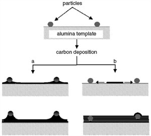
| Possible growth mechanisms for CNTs with particles embedded in the walls: (a) This approach did not work: tip-growth mechanism, with particles lifted from the membrane pore wall due to the formation of a metal carbide particle and further dissolution of carbon in the carbide with subsequent lift of the particle. (b) In the proposed process, carbon first deposits on the alumina and forms a continuous layer due to the faster growth kinetic in the planar direction; eventually, carbon layers lift the particles and then trap them inside the tube walls. (Reprinted with permission from IOP Publishing) | |
| Gogotsi, a professor in the Department of Materials Science and Engineering, heads the Nanomaterials Group and the A.J. Drexel Nanotechnology Institute at Drexel University. His recent paper in Nanotechnology, "Multifunctional carbon nanotubes with nanoparticles embedded in their walls", describes how controlled amounts of nanoparticles ranging in size and composition were embedded in the walls of carbon nanotubes during a one-step, template-assisted chemical vapor deposition (CVD) process. | |
| Gogotsi points out that one key advantage of the tubes prepared in the experiments is that the cavity of the CNT is empty. This is not always the case with tubes whose cavity is filled by magnetic particles. This could allow CNT alignment in the presence of a magnetic field without interfering with fluid flow inside the CNTs. | |
| These magnetically active tubes could also be manipulated through magnetic assembly in micro- and nano-devices, or loaded with drugs, driven to a specific location inside a cell or used for biosensing. | |
| In the magnetic nanotubes synthesized in this work, metal particles are always covered by a carbon layer which does not allow direct chemical reaction or loss of particles in the environment. | |
| One particularly interesting application of this novel fabrication technique is to assemble a nanoprobe with attractive features for biological investigation. | |
| "By inserting magnetic nanoparticles we can manipulate nanotubes using a magnetic field which, unlike electric fields, does not interact with biomolecules" says Gogotsi. "In collaboration with Prof. G. Friedman and Prof. A. Fontecchio from Drexel Electrical and Computer Engineering department, we assembledthese tubes into nanotube-tipped cellular probes. Connecting magnetic carbon nanotubes to glass pipettes for fluid transfer produces a syringe terminated by a strong, yet flexible tube that is about 1,000 times thinner than a human hair. These nanotubes are robust enough to actually inject a cell." | |
| In a different paper, published last week in Applied Physics Letters ("Magnetically assembled carbon nanotube tipped pipettes"), the Drexel researchers describe such a a biological nanoprobe with a magnetic CNT tip that has the ability to transfer fluids. D. Mattia and G. Korneva, who are PhD students in Prof. Gogotsi's group, produced carbon nanotubes by chemical vapor deposition into alumina templates.? J. Freedman (NSF IGERT Fellow working with A. Fontecchio and G. Friedman) assembled them into nanotube-tipped pipettes. | |
 |
|
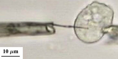
| Optical image of a nanotube tipped pipette (left) is injected into a cell held in place by negative pressure on a patch pipette (right). Negligible deformation of the cell occurs. (Reprinted with permission from American Institute of Physics) | |
| The objective of this probe is to permit molecules of interest to pass into and out of an individual cell, nuclei, or organelle through the conjoined nanotube and pipette device. This allows for controlled substance delivery and quantitative sampling. | |
| Nanotubes with a diameter of about 200 nm were used in these experiments to demonstrate the feasibility of this technique in an optically verifiable regime. Since the size-limiting factor is the magnetic particles (which measure about 10 nm) lining the tubes, the technique can be scaled down to smaller CNTs of 20?40 nm diameter. | |
| "We are now on the verge of single molecule detection inside carbon nanotubes" says Gogotsi. "By combining our nanotubes having gold nanoparticles embedded in the wall with Surface Enhanced Raman Spectroscopy, chemical analysis inside nanotubes can be performed with extreme sensitivity. This technique could enhance biochemical detection capabilities for detection of toxins in air or water." | |
| In a next step, the researchers hope to fully develop nanoscale biomedical probes based on their nanotubes. | |
| "From the materials science point of view, we plan to expand the range of functionalities to be added add to the nanotubes by testing different types of particles, such as quantum dots, for example" says Gogotsi. "We will also attach various molecules to the tube surfaces." | |
 By
Michael
Berger
– Michael is author of three books by the Royal Society of Chemistry:
Nano-Society: Pushing the Boundaries of Technology,
Nanotechnology: The Future is Tiny, and
Nanoengineering: The Skills and Tools Making Technology Invisible
Copyright ©
Nanowerk LLC
By
Michael
Berger
– Michael is author of three books by the Royal Society of Chemistry:
Nano-Society: Pushing the Boundaries of Technology,
Nanotechnology: The Future is Tiny, and
Nanoengineering: The Skills and Tools Making Technology Invisible
Copyright ©
Nanowerk LLC
|
Become a Spotlight guest author! Join our large and growing group of guest contributors. Have you just published a scientific paper or have other exciting developments to share with the nanotechnology community? Here is how to publish on nanowerk.com.