| Posted: Sep 10, 2010 | |
Novel optical procedure greatly facilitates nanotube characterization |
|
| (Nanowerk Spotlight) Single-walled carbon nanotubes (SWCNT) have been on the forefront of nanotechnological research for over a decade due to their outstanding and unique mechanical and electronic properties rendering this novel carbon allotrope an ideal candidate for a variety of electronic applications (see related spotlight articles, for example "A novel single electron pump based on a carbon nanotube" or "DNA-assisted solution processing for high-performance thin-film transistors"). | |
| However, progress towards SWCNT-based technology commenced slowly even though the remarkable potential has been realized soon after their discovery in 1991. | |
| A first major drawback is related to the characterization of functionalized SWCNTs. Since carbon nanotubes are intrinsically insoluble in common organic solvents and water, their surface needs to be modified by covalent or noncovalent functionalization in order to increase their poor processeability. Accordingly, the successful derivatization needs to be analyzed, usually by cumulating evidence from a variety of independent spectroscopic (for example Raman, absorption, NIR emission spectroscopy) and microscopic (for example atomic force microscopy, scanning or transmission electron microscopy) techniques. Thus, a great variety of analytical techniques exists and may be applied to nanotube characterization. | |
| However, it is exactly this diversity that renders nanotube characterization highly challenging, as no standard protocol for the precise analysis has yet been established. Taken as individual methods, every characterization technique has its own limitations and restrictions so that the precise analysis can only be achieved by combining the information from the different techniques. | |
| Our group has recently presented significant progress towards reaching this goal ("Optical Visualization of Carbon Nanotubes – a Unifying Linkage Between Microscopic and Spectroscopic Characterization Techniques"). We have described a readily accessible and low-cost methodology towards correlating spectroscopic and microscopic information by the aid of the optical visualization of one dimensional nano-scaled objects such as SWCNTs when deposited on Si/SiO2 substrates with a SiO2 layer thickness of 300 nm. | |
| Even though imaging of the 2D sibling of SWCNTs, e.g. graphene by optical microscopy on opaque bilayered substrates such as Si/SiO2 has been reported (and widely adopted to facilitate characterization; see for example: "Making graphene visible" and "Soluble Graphene: Soluble Graphene: Generation of Aqueous Graphene Solutions Aided by a Perylenebisimide-Based Bolaamphiphile"), it is quite astonishing that it can be applied to 1D structures as well, as they are below the optical detection limit of λ/2 in two dimensions. | |
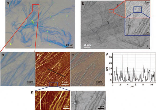
| The protocol to be followed is quite simple, as it involves dispersion of the nanotubes by sonication and deposition (spin coating, drop casting or dip coating) on the magic Si/SiO2 substrates. Upon illumination with normal white incident light in a commercial optical microscope, the structure of the nanotube aggregates appears as interference contrast (Figure 1a). This optical visualization by any optical microscope can then be used to re-localize and analyze exactly the same sample area, for example by atomic force microscopy (figure 1d), scanning electron microscopy (figure 1b) and Raman spectroscopy. Thus, the microscopic information can be correlated to spectroscopic information which is of uttermost importance for the precise investigation by Raman spectroscopy, as the Raman signals of SWCNTs strongly depend on the morphology of the sample. | |
 |
|
| Figure 1. Optical micrographs of SWCNTs spin-casted onto a Si/SiO2 substrate (SiO2 thickness 300 nm). a) Bright field image, b) SEM image with different magnifications of the respective areas, c) enlarged bright field image displaying the same spot as d) atomic force microscopic image; e) overlay of bright field image and AFM image; f) height profile along the black line in d); g) direct comparison of AFM and SEM images from the area indicated by the blue rectangle in figure 1d. (Reprinted with permission from Wiley-VCH Verlag) | |
| The further impact of this optical visualization is presumably striking, as it does not only enable the precise and reliable characterization of SWCNTs by correlative spectroscopy and microscopy, but it may also be used as quick characterization technique itself to estimate the quality of a SWCNT dispersion after deposition on the magic wafers. | |
| Similarly to the graphene sheets in graphite, strong mutual attractive forces between the individual sp2 carbon cylinders glue the SWCNTs together in nanotube bundles rendering pristine SWCNTs virtually insoluble in organic solvents and water without stabilization. This insolubility and non-disperseability can be overcome by dispersion additives such as surfactants and detergents. | |
| However, one needs to distinguish between dispersion of bundled SWCNTs and truly exfoliated and individualized material. The quality of the dispersion, e.g. the degree of SWCNT individualization is commonly characterized by microscopic techniques such as AFM. Since AFM is a scanning technique, a detailed statistical analysis of the sample is time consuming. Accordingly, a quick first estimation of the dispersion quality is highly desirable. | |
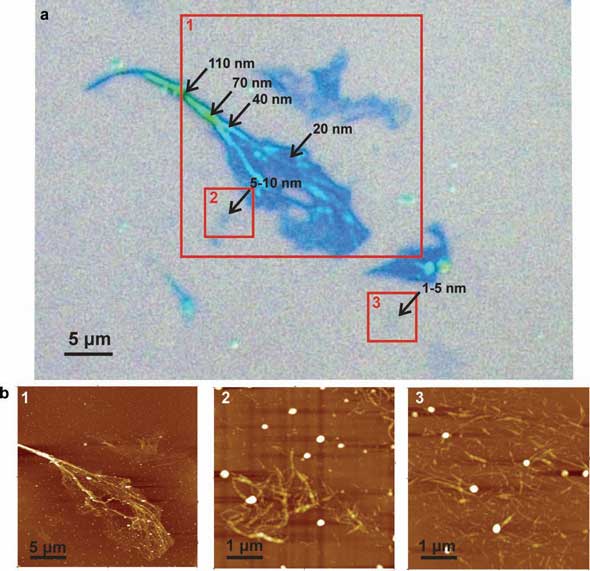
| A tool towards achieving this goal is presented in optical bright field microscopy, as it has been shown that the color of the contrast ranging from deep blue over cyan to green arising from the SWCNTs deposited onto the Si/SiO2 substrates can roughly be correlated to the height of the objects thus yielding a first impression on the dispersion quality (Figure 2). If a detailed analysis by AFM is nonetheless required, the optical visualization also facilitates the AFM analysis, as areas of interest are revealed by the optical contrast so that the wafers can systematically be analyzed. | |
 |
|
| Figure 2. Top: Optical bright field micrograph of SWCNTs spin-casted onto a Si/SiO2 substrate (SiO2 thickness 300 nm), Bottom: AFM tapping mode images corresponding to the areas marked by the red squares. (Reprinted with permission from Wiley-VCH Verlag) | |
| The topic of the SWCNT dispersion quality is closely related to the greatest hurdle in nanotube research, e.g. the polydispersity of the as-produced nanotubes. The SWCNT raw material is intrinsically comprised of a mixture of differing lengths, diameters, chiralities and as a consequence electronic properties, e.g. SWCNTs exhibit metallic or semiconducting characteristics depending on the way the graphene sheet is rolled-up into the cylinder. | |
| Various separation techniques have been proposed and described in the literature such as electrophoresis, chromatography or density gradient ultracentrifugation. Even though nanotube sorting according to diameter or chirality can thus be achieved, the large scale separation of SWCNTs for industrial and commercial applications is a very distant prospect, as only individualized, disaggregated SWCNTs can be separated. | |
| Commercially available surfactants and detergents may overcome the obstacle of SWCNT dispersion. However, the individualization rates are usually poor so that efforts are also directed towards the development of novel SWCNT surfactants capable of not only dispersing, but also of exfoliating SWCNTs to a high degree. Potent candidates for the production of high quality dispersions which may ultimately increase the yields in SWCNT separation and sorting have also recently been developed in our group. For more information see these recent papers: | |
| "Fractioning HiPco and CoMoCAT SWCNTs via density gradient ultracentrifugation by the aid of a novel perylene bisimide derivative surfactant " | |
| "High Population of Individualized SWCNTs through the Adsorption of Water-Soluble Perylenes" | |
| "Nanotube Surfactant Design: The Versatility of Water-Soluble Perylene Bisimides" | |
| "Dispersion of HiPco and CoMoCAT Single-Walled Nanotubes (SWNTs) by Water Soluble Pyrene Derivatives–Depletion of Small Diameter SWNTs" | |
| "Soluble Graphene: Generation of Aqueous Graphene Solutions Aided by a Perylenebisimide-Based Bolaamphiphile" | |
| C. Backes, E. Karabudak, C. D. Schmidt, F. Hauke, A. Hirsch, W. Wohlleben, Chem. Eur. J. 2010, in press, DOI: chem.200903461. | |
| C. Backes, C. D. Schmidt, J. N. Coleman, W. Wohlleben, F. Hauke, A. Hirsch, Chem. Eur. J. 2010, in press, doi:chem.201000232. | |
| By Prof. Dr. Andreas Hirsch and Claudia Backes, Department of Chemistry and Pharmacy & Interdisciplinary Center for Molecular Materials (ICMM) at Friedrich-Alexander-Universität Erlangen-Nürnberg | |
|
Become a Spotlight guest author! Join our large and growing group of guest contributors. Have you just published a scientific paper or have other exciting developments to share with the nanotechnology community? Here is how to publish on nanowerk.com. |
|