| Posted: Jun 26, 2012 | |
Painting with biomolecules at the nanoscale |
|
| (Nanowerk Spotlight) Many nanotechnology projects require some form of nanopatterning technique for fabricating the devices, structures and surfaces required in fields ranging from electronics to photonics, security, biotechnology and medicine. Although they may not be visible to the naked eye, the nanometer-sized trenches, ridges, curves and grooves of these patterns and surfaces have a very visible impact. Nanopatterning is a very delicate task which is only possible with special techniques and methods. | |
| Researchers have developed a wide range of nanopatterning techniques, from top-down methods such as nanoimprint, e-beam or UV lithography to bottom-up techniques such as transfer nanolithography or nanopositioning on DNA or protein scaffolds. | |
| A novel technique, developed by an Austrian research team, uses a biofunctionalization approach based on resist-less electron-beam-induced deposition (EBID) of carbon-containing nanofeatures, that has been developed into a universal biofunctionalization platform. | |
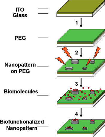
| The team used a high-resolution scanning electron microscope for their EBID patterning. The complete process sequence from e-beam writing to the incubation with biomolecules is schematically summarized below: | |
 |
|
| EBID-process sequence employed for the generation of biomolecular nanopatterns. Following (1) PEG-silanization of the ITO-glass substrate, (2) e-beam-induced deposition generates a pattern of nanosized features. (3) Incubation with biomolecules and (4) rinsing leads to the selective decoration of the nanofeatures with biomolecules. (Reprinted with permission from American Chemical Society) | |
| Reporting their findings in a recent online issue of Nano Letters ("Painting with Biomolecules at the Nanoscale: Biofunctionalization with Tunable Surface Densities"), the team from the Center for Advanced Bioanalysis and the Institute for Biophysics at Johannes Kepler University, describes a nanopatterning strategy for bio-functionalized surfaces, which achieves unprecedented control over several key characteristics. These include an outstandingly high contrast of >1000 between biomolecules bound to the target and nontarget areas, as well as control over three orders of magnitude in the biomolecule densities between different target areas within the same array. | |
| As the scientists note, these remarkable results are based on the combination of four pivotal elements: | |
| "Firstly, the use of high-density PEG films prevents the nonspecific adsorption of biomolecules in the nontarget areas of the array and is thus mainly responsible for the high specificities. Secondly, the electron-beam-induced target areas allow large-range control of the density of adsorbed biomolecules by simply changing the e-beam exposure dose. Thirdly, avidin decoration under optimized pI and pH environments makes this a universal biofunctionalization platform that can be utilized for all biomolecules that bind through a suitable ligand to avidin. And finally, the PEG-ITOcoated glass substrates are fully compatible with highly sensitive optical analysis techniques." | |
| According to the team, "this finding offers the exciting possibility to implement different densities of biomolecules within one and the same nanopattern array." | |
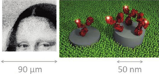
| To demonstrate this ability, the scientists generated a simple gray-scale image of the Mona Lisa using fluorescence-labeled immunoglobulin G (IgG-Cy3). | |
 |
|
| (Left) Fluorescence image of a medieval painting that was implemented as an array of IgG-Cy3 molecules specifically bound to EBID features. Four different levels of brightness were obtained by varying the surface density of the biomolecules via the employed ebeam dose. The area of the fluorescence image is 90 ? 90 µm2 with a pixel spacing of 500 nm, and a pixel diameter of <50 nm. (Right) Schematic view on the pixel level illustrating that the density of IgGCy3 molecules changes with the height of the EBID features. Pixel size and spacing are not to scale. (Reprinted with permission from American Chemical Society) | |
| The resulting fluorescence image in the figure above vividly demonstrates the envisaged density control of the biomolecules within an array, a feature that cannot easily be achieved with other nanopatterning strategies. | |
| "We expect that this unique ability can be exploited for biological experiments, where cells respond to the nanoscale density of activating molecules such as antibodies" write the authors. | |
 By
Michael
Berger
– Michael is author of three books by the Royal Society of Chemistry:
Nano-Society: Pushing the Boundaries of Technology,
Nanotechnology: The Future is Tiny, and
Nanoengineering: The Skills and Tools Making Technology Invisible
Copyright ©
Nanowerk LLC
By
Michael
Berger
– Michael is author of three books by the Royal Society of Chemistry:
Nano-Society: Pushing the Boundaries of Technology,
Nanotechnology: The Future is Tiny, and
Nanoengineering: The Skills and Tools Making Technology Invisible
Copyright ©
Nanowerk LLC
|
|
|
Become a Spotlight guest author! Join our large and growing group of guest contributors. Have you just published a scientific paper or have other exciting developments to share with the nanotechnology community? Here is how to publish on nanowerk.com. |
|