| Posted: Oct 15, 2007 | |
Quantum dot imaging could benefit embryonic stem cell therapy |
|
| (Nanowerk Spotlight) A quantum dot (QD), also called a nanocrystal, is a semiconductor nanostructure that can be as small as 2 to 10 nm. The usefulness of quantum dots comes from their peak emission frequency's extreme sensitivity - quantum mechanical in nature - to both the dot's size and composition. QDs have been touted as possible replacements for organic dyes in the imaging of biological systems, due to their excellent fluorescent properties, good chemical stability, broad excitation ranges and high photobleaching thresholds. By contrast, conventional organic dyes cannot be easily synthesized to emit different colors and have narrow excitation spectra and broad emission spectra that often cross into the red wavelengths, making it difficult to use these dyes for multiplexing. QDs hold increasing potential for cellular imaging both in vitro and in vivo. Researchers have now used QDs for in vivo imaging of embryonic stem cells in mice. This opens up the possibility of using QDs for fast and accurate imaging applications in stem cell therapy. | |
| Stem cell therapy is the process of injecting stem cells into an organism in the hope that they will differentiate and replace damaged tissue or grow new organs. This technology holds great promise for treatment of a wide range of intractable conditions such as Parkinson’s disease, diabetes, or degenerative joint diseases. There are two types of cells used in stem cell therapy, adult stem cells and embryonic stem (ES) cells. ES cells are the ultimate source for use in cell-based therapy because they posses a virtually unlimited capacity for self-renewal and because they possess the ability to differentiate into all other cell types found in the body, from brain cell to toe nail. For stem cell therapy, it is important to develop methods to monitor cell survival and location after transplantation. | |
| "In stem cell therapy, monitoring of cell survival and location after transplantation is important for determining their efficacy" Dr. Joseph C. Wu explains to Nanowerk. "Because the absorption and scattering of light in biological tissue can be considerable, any optical signal transmitted from deep tissues to the surface tends to diminish in strength. With QDs’ many advantages over traditional organic dyes, QDs may provide an excellent tool for imaging stem cell therapy." | |
 |
|
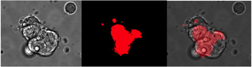
| Fluorescent images of embryonic mouse stem cells labeled with QDs on day 1 post labeling. (Copyright: BioMed Central) | |
| Wu, an Assistant Professor of Medicine & Radiology at Stanford University School of Medicine, together with collaborators from the University's Molecular Imaging Program, conducted a study in which the scientists used the peptide-based reagent QTracker to label mouse ES cells with QDs and evaluate the utility of QDs for imaging stem cell therapy." The results have been published in a recent free access paper in the July issue of BMC Biotechnology ("Quantum dot imaging for embryonic stem cells") | |
| Wu and his colleagues successfully labeled murine embryonic stem cells with six different quantum dots and demonstrated the ES cell viability, proliferation, and differentiation were not adversely affected by QDs. They showed that QD 525, QD 565, QD605, QD 655, QD 705, and QD 800 labeled ES cells can be detected in vivo using a single excitation wavelength (465 nm). This finding makes QDs an attractive choice for regenerative therapy. | |
 |
|
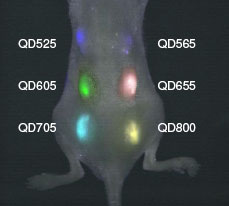
| 1 million ES cells labeled with QD 525, 565, 605, 655, 705, and 800 were subcutaneously injected on the back of the athymic nude mice right after labeling and the image was taken with a single excitation light source right after injection. (Copyright: BioMed Central) | |
| "We also successfully imaged labeled ES cells with good contrast with one single excitation wavelength in vivo" says Wu. "This versatility makes them good candidates for tumor targeting, lymph node and vascular mapping, and cell trafficking in small animal imaging." | |
| Toxicity of QDs of course is a key factor in determining whether it will be a feasible probe for both cellular and clinical use. The Stanford scientists carefully examined QDs’ effect on ES cells and found that QD labeling had no detectable effect on ES cell growth. Next they tested its effect on cellular development and differentiation and again found no effects of the labeling. | |
| Wu points out that another advantage of QDs is their ability to do multiplex imaging of different QDs at the same time. "However" he says, "in our study, ES cells labeled with different QDs were only capable of being imaged up to day 2 after subcutaneous implantation. A likely cause for this could be the loss of signal due to rapid cell division. Another possible cause could be serum instability of the QDs." | |
| While this study appears to be the first successful demonstration of in vivo multiplex imaging of mouse ES cells labeled QDs, the use of QDs in stem cells is only beginning to be explored. | |
| Due to their many advantages over conventional organic dyes, QDs serve as good candidates to monitor cell survival and location after transplantation in stem cell therapy However, the poor retention of QDs in targets cells may be a problem for long-term tracking. Wu says that upon further improvements – such as near-infrared QDs, better serum stability, and improved cell retention – QDs will have greater potential for tracking of stem cells within deep tissues. | |
 By
Michael
Berger
– Michael is author of three books by the Royal Society of Chemistry:
Nano-Society: Pushing the Boundaries of Technology,
Nanotechnology: The Future is Tiny, and
Nanoengineering: The Skills and Tools Making Technology Invisible
Copyright ©
Nanowerk LLC
By
Michael
Berger
– Michael is author of three books by the Royal Society of Chemistry:
Nano-Society: Pushing the Boundaries of Technology,
Nanotechnology: The Future is Tiny, and
Nanoengineering: The Skills and Tools Making Technology Invisible
Copyright ©
Nanowerk LLC
|
|
|
Become a Spotlight guest author! Join our large and growing group of guest contributors. Have you just published a scientific paper or have other exciting developments to share with the nanotechnology community? Here is how to publish on nanowerk.com. |
|