| Posted: Feb 26, 2009 | |
Improved nanotechnology implants through nanopatterned metal surfaces |
|
| (Nanowerk Spotlight) In the quest to make bone, joint and tooth implants almost as good as nature's own version, scientists are turning to nanotechnology. Researchers have found that the response of host organisms (including at the protein and cellular level) to nanomaterials is different than that observed to conventional materials. While this new field of nanomedical implants is in its very early stage, it holds the promise of novel and improved implant materials. | |
| One recent example is the nanopatterning of metal surfaces that promises to lead to superior medical implants. A multidisciplinary team of scientists have demonstrated that a simple and inexpensive chemical treatment can create nanopatterns on the surface of different implantable metals, such as Titanium, Tantalum, and CrCoMo alloys. | |
| "We found that, by immersing these metals in an etching solution made by mixing an acid (or base) and an oxidant, it is possible to create a variety of reproducible sponge-like networks of nanopits on the material's surface" Fiorenzo Vetrone tells Nanowerk. "We can now precisely engineer the nanoscale physicochemical characteristics of surfaces by selecting the appropriate etching solution. We have shown that the resulting nanopatterned surfaces have significant effects on different cellular events in vitro, which conferred novel biological functionalities to the implantable metals." | |
| "Our study is groundbreaking," adds Antonio Nanci. "We use simple yet very efficient chemical treatments to alter metals commonly used in the operating room. This innovative approach may ultimately hold the key to developing intelligent materials that are not only easily accepted by the human body but that can actively respond to the surrounding biological environment." | |
 |
|
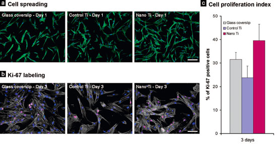
| Human umbilical cord (HUC)-derived cells grown on control Ti surfaces, nanotextured Ti surfaces, and on control glass coverslips. (a) At day 1, HUC cells were well spread on all surfaces, exhibiting an elongated shape. Areas of higher cell density were frequently observed for nanostructured Ti. (b, c) Dual nuclear labeling with an anti-Ki-67 antibody (red fluorescence) and DAPI (blue fluorescence) at day 3 allowed the detection of significantly more cycling cells on nanotextured Ti compared to controls (1.6-fold increase compared to control Ti). Phalloidin labeling appears green in (a) and pale white in (b). Scale bar ) 200 µm (a) and 100 µm (b). (reprinted with permission from American Chemical Society) | |
| Nanci and Vetrone are members of a team of scientists from the Université de Montréal, McGill University, the Institut National de la Recherche Scientifique (INRS-EMT), Plasmionique Inc and the Universidade de São Paulo. Their findings, first authored by Vetrone, have been published in a recent paper in Nano Letters (Nanoscale Oxidative Patterning of Metallic Surfaces to Modulate Cell Activity and Fate). | |
| Earlier work on improving the biocompatibility of medical implant materials with nanotechnology has focused on nanoscale bumps, protrusions, and other features created by diverse techniques that are proven to have highly promising effects on the activity of cells, which are known to respond by modulating their adhesion, proliferation, migration, and gene expression. However, the majority of these nanoscale features were created with expensive and cumbersome instrumentation. | |
| In contrast, Nanci's team uses a simple chemical treatment to create nanopatterns on the surface of various biocompatible metals that is amenable to production of nanoscale features on three-dimensional prosthetic devices. | |
| These biocompatible metals can now actively interact with the surrounding biological environment and deliver specific signals to guide and control cell activities without the addition of exogenous chemical agents (i.e. growth factors, drugs). | |
| Nanci says that the results prove that chemical oxidation is a simple yet powerful tool to direct a priori cell-material interactions towards a specific pathway. "For example, these nanostructured surfaces have demonstrated the ability to guide stem cells towards the osteogenic pathway. Furthermore, we are now able to endow implantable metals with the capacity to selectively promote or limit the adhesion and growth of different cells." | |
| "The factors, which prompted our research, coincide with our goals," says Vetrone: "the necessity to extend our knowledge of cellsubstrate interactions. In the human body, cellular events occur at the implant-host tissue interface and they are regulated by the surface properties of the materials. Understanding how cells respond to a foreign surface will contribute contemporaneously to the progress of biological sciences and that of health-related research. In fact, unraveling the processes, which control cell activities is fundamental to achieve an improved comprehension of the cellular and molecular biology, as well as a novel generation of implantable materials able to positively interact with the surrounding tissues." | |
| Potential applications of this work range from in vitro studies of cell-substrate interactions to nanomedicine. Chemically generated nanopatterns can be exploited for detailed studies of how cells respond to nanometric cues. In addition, considering the ease of exporting the chemical treatment to a large-scale production facility, it will be possible to easily create nanopatterned implants for dental, orthopedic and cardiovascular applications with improved biological responses. | |
| For example, orthopedic and dental implants will be able to accelerate osseo-integration and prosthesis fixation by enhancing bone growth and regeneration while limiting that of connective tissues. Another possible application are cardiovascular stents that have anticoagulant properties, which will avoid thrombosis, as well as the capacity to control drug delivery at the implantation site. | |
| One caveat of these findings is that most of the experiments have been carried out in cell culture but these in vitro conditions cannot accurately represent the complex biochemical system of living entities. The scientists therefore aim to evaluate the relevance of nanopatterned surfaces in vivo, to determine whether the enhanced biological functions of the modified materials are conserved. | |
| The major conclusion of these studies though is that the beneficial influence of microtopography on osteogenic cell activity actually results from chemical modification of the surface rather than from topographic cuing. | |
| "It must also be noted that surface modifications also affect the adsorption of proteins, which then secondarily alters cellular activity," says Vetrone. | |
 By
Michael
Berger
– Michael is author of three books by the Royal Society of Chemistry:
Nano-Society: Pushing the Boundaries of Technology,
Nanotechnology: The Future is Tiny, and
Nanoengineering: The Skills and Tools Making Technology Invisible
Copyright ©
Nanowerk LLC
By
Michael
Berger
– Michael is author of three books by the Royal Society of Chemistry:
Nano-Society: Pushing the Boundaries of Technology,
Nanotechnology: The Future is Tiny, and
Nanoengineering: The Skills and Tools Making Technology Invisible
Copyright ©
Nanowerk LLC
|
|
|
Become a Spotlight guest author! Join our large and growing group of guest contributors. Have you just published a scientific paper or have other exciting developments to share with the nanotechnology community? Here is how to publish on nanowerk.com. |
|