Showing Spotlights 409 - 416 of 544 in category All (newest first):
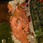 Bone grafts are second only to blood transfusions on the list of transplanted materials. With bone grafts, a surgeon replaces missing bone with material from the patient's own body, or a synthetic or natural substitute. Traditional tissue repair techniques, especially with synthetic materials, may lead to poor integration with the existing bone or tissue structure, potentially transfers dangerous pathogens into the body, and sometimes lead to a complete rejection of the graft. To minimize these harmful side effects, researchers are developing sophisticated tissue engineering techniques based on the construction of three-dimensional scaffolds out of biomaterials to provide mechanical support and guide cell growth into new tissues or organs. In the quest to make bone, joint and tooth implants almost as good as nature's own version, scientists are turning to nanotechnology. They have found that the response of host organisms to nanomaterials is different than that observed with conventional materials. The surface nano-characteristics of biomaterials are increasingly recognized as crucial factors with regard to tissue acceptance and cell behaviors. While this new field of nanomedical implants is in its very early stage, it holds the promise of novel and improved implant materials.
Nov 4th, 2008
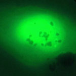
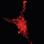 The study of individual live cells is a hugely important scientific task and essential to the field of molecular biology and biomedical research. Among the most significant technical challenges for performing successful live-cell imaging experiments is to maintain the cells in a healthy state and functioning normally on the microscope stage while being illuminated. Especially if scientists want to look into cellular processes that occur inside cells in their natural state and that cannot be observed by traditional cytological methods. Quantum dots (QDs), also called nanocrystals, hold increasing potential for in vitro and in vivo cellular imaging. For instance, we have previously reported about how researchers have used QDs for in vivo imaging of embryonic stem cells in mice, a novel technique that has opened up the possibility of using QDs for fast and accurate imaging applications in stem cell therapy. The usefulness of quantum dots comes from their peak emission frequency's extreme sensitivity to both the dot's size and composition. QDs have been touted as possible replacements for organic dyes in the imaging of biological systems, due to their excellent fluorescent properties, good chemical stability, broad excitation ranges and high photobleaching thresholds.
Oct 8th, 2008
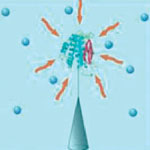 Genomics and proteomics, the studies of genes and proteins, provide the underlying basis for many advances in drug development and effective treatments of diseases. These studies heavily rely on unveiling the behavior of a single DNA or protein in an investigative sample. You could compare this challenge to somehow finding, then catching and monitoring a particular fish in a vast ocean. The scientific term for 'catching the fish' is 'immobilization' - a powerful technique for the study of biochemical systems that allows for the continuous observation of dynamic behavior of a chosen target. Immobilization methods anchor the to be observed molecule onto a surface in order to restrict it from escaping the observation volume. Researchers have now developed a new platform which consists of a carbon nanotube nanoneedle for capturing, isolating and measuring the activity of miniscule amounts of proteins.
Oct 3rd, 2008
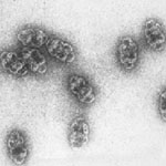 Spheres can be found at all scales in both the inanimate and living world for the basic physical property of encapsulation. There has been a fair amount of work by nanotechnology researchers on putting non-biological molecules or clusters into viruses or virus-like artificial nanocontainers. Although viruses are a type of protein cage, they are not a natural part of the cell. There is a class of biological protein nanocapsules though - called vaults - that is part of cells - although their cellular functions and gating mechanism are not yet understood. Vault nanoparticles are already present in human cells in high numbers (approx. 10,000 per cell) and their hollow barrel-like structure with a large internal volume seems well suited for encapsulation purposes.
Oct 2nd, 2008
 Radio frequency ablation (RF ablation) is a treatment for cancer that works by inserting a thin needle guided by computed tomography or ultrasound through the skin and into a tumor. Electrical energy is then delivered through a number of electrodes deployed through the needle, causing a zone of thermal destruction that encompasses the tumor. Researchers have experimented with non invasive radiowave thermal ablation of cancer cells that uses nanoparticles as a novel approach to treat cancer. The idea is that RF treatment of malignant tumors at any site in the body should be possible if it were possible to get agents that release heat under the influence of the RF field to the specific tumor site. Researchers have now developed a novel nanomaterial that has proven to be a very strong RF absorber and provides high enough thermal-ablation ability in order to generate localized thermally-driven damages inside the cancer cells. Even more, these nanoparticles have shown relatively low cytotoxicity and they absorb low frequency RF radiation, which has significant penetration depth inside living organism.
Sep 24th, 2008
 Using nanoparticles to combat cancer and a host of other diseases is an active area of research for many scientists. For decades, treating cancer has mostly involved injecting patients with toxic drugs. A practice, in which only a fraction of the drugs reach the intended target, killing healthy cells in the process and causing harmful side effects. Previous studies have shown that drug-carrying nanoparticles can accumulate in and attack tumors, in part because of their extremely small size, which helps allow them to pass through cell membranes. However, up until now, existing techniques have meant that targeting agents could only be delivered using spherical or granular shaped particles. Now, a team of scientists have demonstrated that nanoparticles designed with a specific shape, size and surface chemistry are taken up into cells and behave differently within cells depending on the characteristics of the particle.
Sep 11th, 2008
 Several aspects determine how effective a pharmaceutical drug is. Probably the key issue is how well a drug molecule is able to reach its intended target. This need for target specific delivery of drugs has been well accepted in modern drug therapy. Many research efforts are geared towards improving not just the tissue accumulation, but also the cell-specific accumulation of drug molecules in the hope of improving the efficacy of these drug molecules. The ability to target nanoparticles to specific types of cancer cells is one of the main reasons that nanoparticles have gained favor as a promising drug delivery vehicle. By increasing the amount of an anticancer agent that gets to tumor cells, as opposed to healthy cells, researchers hope to minimize the potential side effects of therapy while maximizing therapeutic response. Now, a group of scientists has taken this approach one step farther by targeting the specific location inside a tumor cell, where a cancer drug then exerts its cell-killing activity.
Sep 5th, 2008
 Nanomedicine, especially drug delivery with nano-sized drug carriers, is all the rage these days. The concept sounds simple: make nanoscale containers that can escape detection by the body's defense mechanisms, fill them with a drug, get them to the desired location within the body, release the drug payload and, presto, you've got a very effective and efficient weapon for instance to fight cancer. That this model works in principle has already been demonstrated in numerous studies. The same studies show the complicated nature and the many difficulties that scientists are facing in fabricating the right nanocontainers, getting them to the right location, controlling the release mechanism of the drug, measuring the drugs' efficacy, and monitoring the now empty delivery vehicles' fate. Researchers from the Indian Institute of Technology Guwahati present experimental results which suggest that the specificity of release of encapsulated nanoparticles could be achieved with an appropriate combination of encapsulating materials and the choice of an appropriate enzyme that would cleave the encapsulation to release the nanoparticles.
Aug 28th, 2008
 Subscribe to our Nanotechnology Spotlight feed