| Apr 19, 2018 |
Cancer drug observed at work
|
|
(Nanowerk News) Using a Raman microscope, researchers at Ruhr-Universität Bochum (RUB) have studied at which targets the cancer drug Neratinib binds in cells and how its chemical structure changes. Compared with other techniques, this method offers a considerable advantage, as it is not necessary to apply a label to the drug that would indicate its distribution indirectly; rather, the drug itself can be monitored.
|
|
The team headed by Prof Dr Klaus Gerwert and Dr Samir El-Mashtoly from the RUB Department of Biophysics published a report on their work in the journal Angewandte Chemie ("Raman Micro-Spectroscopic Evidence for the Metabolism of a Tyrosine Kinase Inhibitor, Neratinib, in Cancer Cells").
|
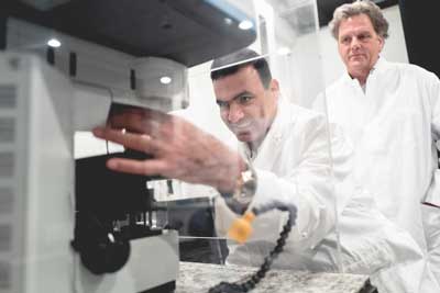 |
| The methods developed by Samir El-Mashtoly (on the left) and Klaus Gerwert are interesting for pharmaceutical companys. (Image: RUB)
|
Pharmacokinetics is crucial
|
|
Neratinib, available under the trade name Nerlynx, has been approved for the treatment of breast cancer since 2017 and is currently in clinical trials for application in lung cancer therapy. It binds to specific proteins in the cell, and thus inhibits cell growth.
|
|
“To understand how a drug works, its pharmacokinetics has to be understood, i.e. how the body absorbs, distributes, and metabolises the drug – as well as how the drug chemically changes,” says Gerwert.
|
How metabolism changes the drug
|
|
In the recent years, Samir El-Mashtoly and Klaus Gerwert have developed novel methods of Raman spectroscopy in order to monitor the efficacy and distribution of drugs in cancer cells. In the current study, the researchers tracked how Neratinib is absorbed in the cells and accumulated in cell organelles. In the cell, the structure of drug molecules changed.
|
|
“Interestingly, most of the drug molecules adopt a structure that is no longer pharmacologically active,” describes El-Mashtoly.
|
|
“These findings can help advance drug development and improve cancer therapy,” he adds. “The study has demonstrated the great potential of Raman microscopy for understanding the mechanisms of action of drugs. Already, we have created a stir in the pharmaceutical industry.”
|
Label-free method
|
|
The common approach for monitoring the drug localisation in cells is to use fluorescent-labelled drug molecules. “Those labels are often larger than the drug molecule itself and thus strongly influence its behaviour,” explains Klaus Gerwert. “The label-free approach is the only technique that enables us to observe the drug directly.”
|
|
Raman microscopy is a label-free method with a spatial resolution of approximately 400 nanometres. In addition to detecting individual cancer cells, it allows visualizing cellular components. Unlike conventional methods, Raman microscopy can detect the chemical changes of drugs that are crucial for their efficacy..
|
|
A pharmaceutical company has become aware of the Bochum-based researchers’ work and uses the method for testing potential cancer drugs.
|