| Posted: | |
| (Nanowerk Spotlight – Application Note) Last year, NT-MDT Co. invited its AFM probe users to take part in an open contest of images obtained with NT-MDT probes. | |
| Though equipment is important in gathering a great AFM image, the result also depends on the probe being used. The purpose of the contest was to compile the most intriguing images collected from ventures into the nano-universe with NT-MDT tips. | |
| Another aim of the ProIMAGE Contest was to show a great variety of scientific and artistic results obtained with a wide range of specialized probes. | |
| A gallery of all images submitted to the contest, which now is closed, and the six winners can be seen on the ProIMAGE webpages. Here is a random selection of some visually stunning images from the 150 contest submissions. | |
 |
|
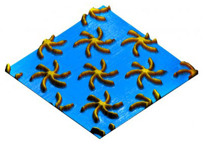
| Micro Starfish. Copper particles on silicon substrate. Fabricated by SEM lithography and ion etching process. Author: Dr. Boris A. Gribkov, Institute for Physics of Microstructures RAS. Scan size: 5x5 µm. Probe used: NSG11. | |
 |
|
| Inverted microscopy. A probe tip was deposited with nanoparticles. Reverse imaging of deposited material was achieved by scanning over a characterization silicon grating TGT (Inverted AFM). This ensures easy localization of deposited nanoparticles, with nanometric accuracy. Author: Dr. Marinello Francesco, DIMEG - University of Padova. Scan size: 5x5 µm. Probe used: NSG03. | |
 |
|
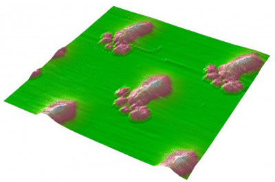
| Wrinkles. Substrate: low resistivity silicon wafer; PDMS (a silicone rubber) on top of the substrate; 5nm sputtered titanium on top of the PDMS. Author: Miss Ruoxuan Li, DIMES, Delft Institute of Microsystems and nanoElectronicS. Scan size: 10x10 µm. Probe used: NSG10. | |
 |
|
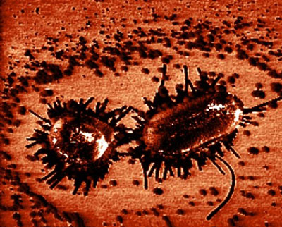
| Escherichia coli sex. Echerichia coli exchanging genetic material. Bcateria were grown in normal LB paltes at standard condition. Author: Kristina Elersic, Institut Jozef Stefan. Scan size: 7x7 µm. Probe used: CSG10/Pt. | |
 |
|
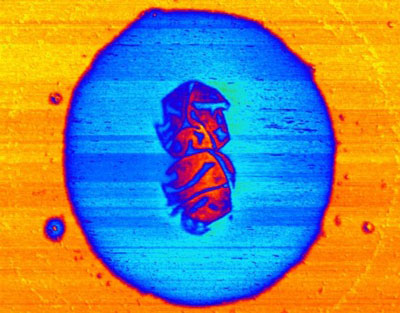
| Capsulated bacteria. A capsulated dividing bacterium was imaged in phase mode in air. The difference of viscoelasticity between the bacteria and the surrounding capsule gave rise to the contrast and the fine details on the surface of the dividing bacterium was revealed in phase mode. Author: Dr. Li Ang. National University of Singapore. Scan size: 3x3 µm. Probe used: NSG11. | |
 |
|
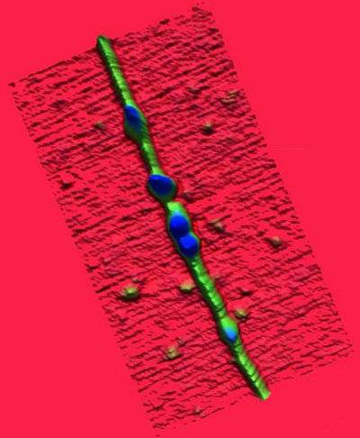
| Single protein, single carbon nanotube. AFM image of complex: single protein (Trypsin) - single wall carbon nanotube deposited on mica. These biomolecules (radius of gyration ∼6nm) adsorb spontaneously on the sidewalls of SWNT. The atomically flat surface is required in order to observe SWNT with diameter ∼1-1.7 nm. There is no binding of CNT on negatively charge surface of mica in absence of biomolecules. The coupling of carbon nanotubes (CNTs) with biomolecules represents a prototype system for bio-nanomaterials conjugates, which preludes to the integration of electronic functionality into biomolecular recognition. Author: Dr. Eva Bystrenova, ISMN-CNR. Scan size: 80x250 nm. Probe used: NSG11. | |
 By
Michael
Berger
– Michael is author of three books by the Royal Society of Chemistry:
Nano-Society: Pushing the Boundaries of Technology,
Nanotechnology: The Future is Tiny, and
Nanoengineering: The Skills and Tools Making Technology Invisible
Copyright ©
Nanowerk LLC
By
Michael
Berger
– Michael is author of three books by the Royal Society of Chemistry:
Nano-Society: Pushing the Boundaries of Technology,
Nanotechnology: The Future is Tiny, and
Nanoengineering: The Skills and Tools Making Technology Invisible
Copyright ©
Nanowerk LLC
|
|
|
Become a Spotlight guest author! Join our large and growing group of guest contributors. Have you just published a scientific paper or have other exciting developments to share with the nanotechnology community? Here is how to publish on nanowerk.com. |
|